. 2003 Aug;163(2):621-32.
doi: 10.1016/S0002-9440(10)63689-9.

Hepatocyte growth factor suppresses renal interstitial myofibroblast activation and intercepts Smad signal transduction

Affiliations

Hepatocyte growth factor suppresses renal interstitial myofibroblast activation and intercepts Smad signal transduction

Junwei Yang et al. Am J Pathol. 2003 Aug.

Abstract

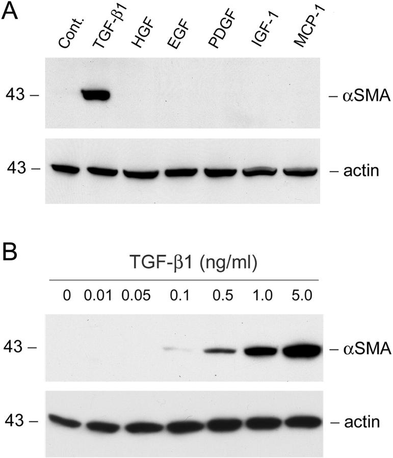
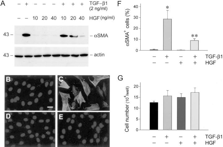
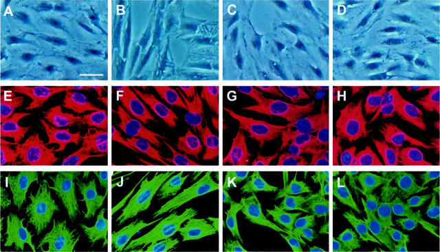
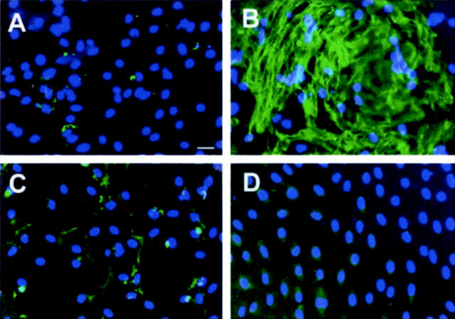
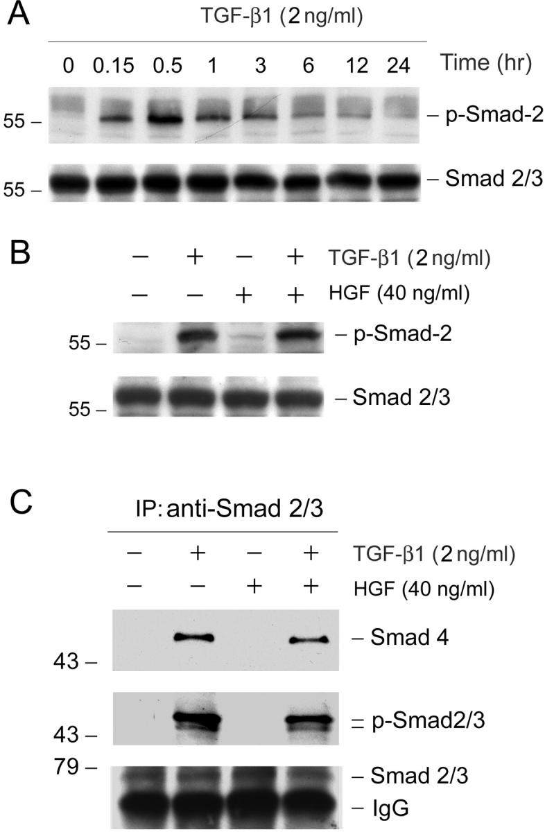
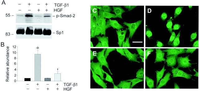
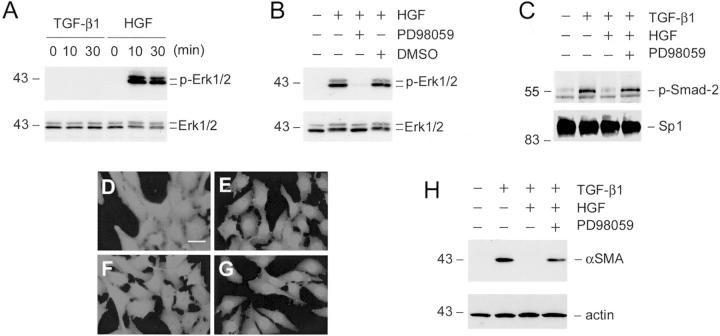
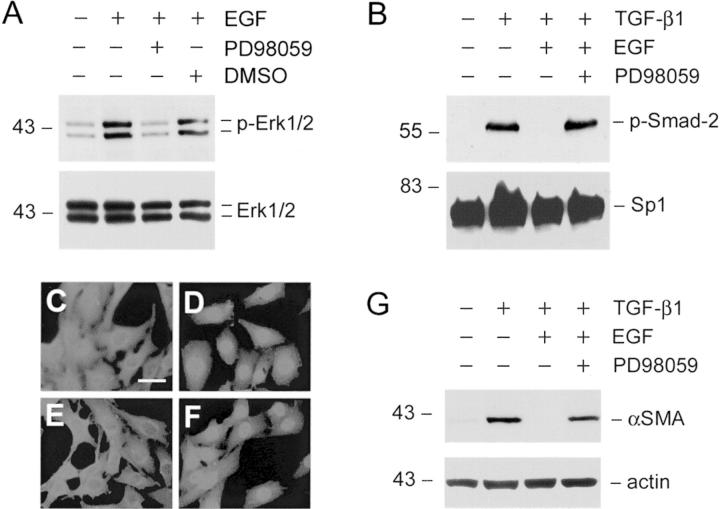
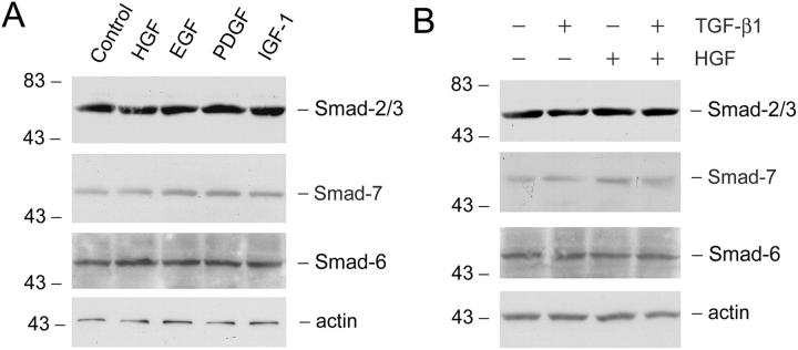
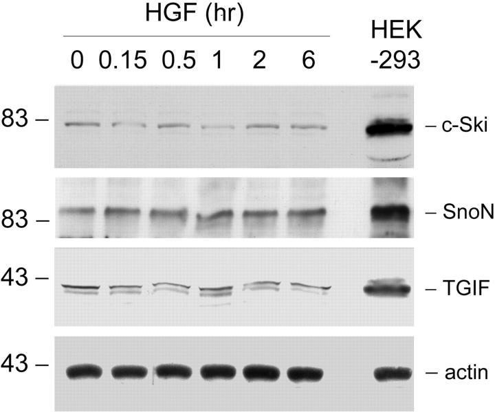
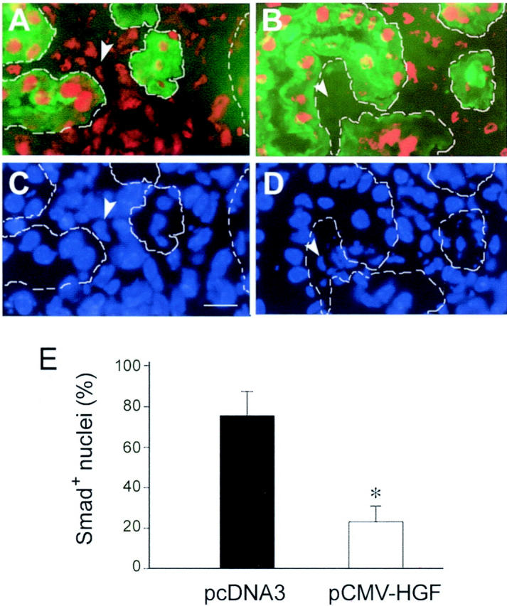
Interstitial myofibroblasts are alpha-smooth muscle actin-positive cells that play a crucial role in the accumulation of excess extracellular matrix during renal interstitial fibrogenesis. Despite their importance in the pathogenesis of renal fibrosis, relatively little is known about the regulators and the mechanism controlling the activation of renal interstitial myofibroblasts in disease conditions. Here, we show that hepatocyte growth factor (HGF) acts as a potent inhibitor of the transforming growth factor (TGF)-beta1-mediated myofibroblastic activation from normal rat renal interstitial fibroblasts (NRK-49F). Simultaneous incubation of HGF abolished TGF-beta1-induced de novo alpha-smooth muscle actin expression, F-actin reorganization, and interstitial collagen I overproduction in a dose-dependent manner. To decipher the mechanism underlying HGF antagonizing TGF-beta1's action, we examined the effects of HGF on TGF-beta1-mediated Smad signaling. HGF neither inhibited Smad-2/3 phosphorylation and their association with Smad-4 induced by TGF-beta1, nor significantly affected inhibitory Smad-6 and -7 expression and cellular abundance of Smad transcriptional co-repressors in NRK-49F cells. However, pretreatment with HGF markedly attenuated activated Smad-2/3 nuclear translocation and accumulation. This action of HGF was apparently dependent on HGF-mediated extracellular signal-regulated kinase-1 and -2 (Erk-1/2) phosphorylation and activation. Inhibition of Erk-1/2 activation by Mek kinase inhibitor PD98059 restored TGF-beta1-mediated Smad-2/3 nuclear accumulation and myofibroblast activation. In vivo, HGF selectively blocked Smad-2/3 nuclear accumulation in renal interstitial cells in the fibrotic kidneys induced by unilateral ureteral obstruction. Therefore, HGF suppresses TGF-beta1-mediated renal interstitial myofibroblastic activation; and this action of HGF is likely related to a mitogen-activated protein kinase-dependent blockade of Smad nuclear translocation.


Figures

Figure 1.
Figure 1.
TGF-β1 induces de novo expression of α-SMA in renal interstitial fibroblast NRK-49F cells. A: NRK-49F cells were incubated for 2 days without (control) or with the same molar concentration (0.2 nmol/L) of various cytokines. The cell lysates were immunoblotted with specific antibodies against α-SMA and actin, respectively. B: Dose-dependent induction of α-SMA expression by TGF-β1 in NRK-49F cells. The cells were incubated with TGF-β1 at the specific concentrations as indicated for 2 days. The α-SMA expression was detected by Western blot analysis.
Figure 2.
Figure 2.
HGF abrogates the expression of α-SMA in renal interstitial fibroblast NRK-49F cells. A: Western blot analysis demonstrates that HGF blocked α-SMA expression induced by TGF-β1 in a dose-dependent manner in NRK-49F cells. Cells were incubated with a fixed amount of TGF-β1 (2 ng/ml) and increasing amounts of HGF as indicated for 2 days. The cell lysates were probed with antibodies against α-SMA and actin, respectively. B to E: Representative photographs of the α-SMA visualized by indirect immunofluorescence staining in NRK-49F cells after various treatments for 2 days. B: Control; C: 2 ng/ml of TGF-β1; D: 40 ng/ml of HGF; E: TGF-β1 plus HGF. C: The α-SMA-positive microfilaments were evident in TGF-β1-treated cells. F: Graphic presentation of the percentage of α-SMA-positive cells in various groups. *, P < 0.01 versus control; **, P < 0.05 versus TGF-β1 group (n = 3). G: Neither TGF-β1 nor HGF significantly affected renal interstitial fibroblast cell growth. NRK-49F cells were treated with 2 ng/ml of TGF-β1, 40 ng/ml of HGF, or both in serum-free medium for 3 days. Cell numbers were counted and presented as means ± SE. No statistically significant difference in cell numbers was found in various groups (n = 3). Scale bar, 10 μm.
Figure 3.
Figure 3.
HGF blocks TGF-β1-induced morphological transformation, F-actin, and vimentin reorganization in renal interstitial fibroblast NRK-49F cells. NRK-49F cells were incubated without (control) (A, E, I) or with 2 ng/ml of TGF-β1 (B, F, J), 40 ng/ml of HGF (C, G, K), or both (D, H, L) for 2 days. A to D: TGF-β1 induced morphological transformation of NRK-49F cells into a myofibroblastic appearance (B). HGF blocked this transformation (D). E to L: Representative micrographs of tetramethyl-rhodamine isothiocyanate-conjugated phalloidin staining (E–H) and vimentin staining (I–L) showing F-actin and vimentin reorganization in NRK-49F cells induced by TGF-β1 (F, J). HGF primarily abolished TGF-β1-induced actin and vimentin reorganization (H, L). Scale bar, 20 μm.
Figure 4.
Figure 4.
HGF suppresses TGF-β1-induced collagen I expression and its extracellular assembly in renal interstitial fibroblast NRK-49F cells. Immunofluorescence staining shows the distribution and abundance of collagen I in NRK-49F cells after various treatments. A: Control; B: 2 ng/ml of TGF-β1; C: 40 ng/ml of HGF; D: TGF-β1 plus HGF. B: TGF-β1 induced interstitial matrix component collagen I expression and its extracellular assembly. D: HGF abrogated TGF-β1-initiated collagen I expression and extracellular assembly. Scale bar, 10 μm.
Figure 5.
Figure 5.
HGF neither inhibits TGF-β1-initiated Smad-2/3 phosphorylation nor affects their association of Smad-4 in renal interstitial fibroblast NRK-49F cells. A: Kinetics of Smad-2 phosphorylation and activation after TGF-β1 incubation in NRK-49F cells. Cells were treated with 2 ng/ml of TGF-β1 for various periods of time as indicated. The cell lysates were probed with antibodies against phospho-specific Smad-2 and total Smad-2, respectively. B: HGF did not inhibit Smad-2 phosphorylation triggered by TGF-β1. Cells were pretreated with 40 ng/ml of HGF for 30 minutes and followed by incubation with 2 ng/ml of TGF-β1 for an additional 30 minutes. Activation of Smad-2 was assessed by using phospho-specific Smad-2 antibody. C: HGF does not affect Smad-2/3 phosphorylation and their association with Smad-4 induced by TGF-β1. NRK-49F cells were pretreated with 40 ng/ml of HGF for 30 minutes and followed by incubation with 2 ng/ml of TGF-β1 for an additional 30 minutes. Cell lysates were immunoprecipitated with antibody against Smad-2/3. The precipitated complexes were immunoblotted with antibodies against Smad-4, phosphoserine, and Smad-2/3, respectively.
Figure 6.
Figure 6.
HGF blocks activated Smad-2 nuclear translocation and accumulation in renal interstitial fibroblast NRK-49F cells. A: Representative Western blot demonstrates that HGF attenuated nuclear accumulation of activated Smad-2 triggered by TGF-β1. Cell nuclei were isolated from NRK-49F cells after various treatments as indicated and nuclear accumulation of phospho-Smad-2 was examined by Western blot. The samples were also probed with ubiquitous transcription factor Sp1 for normalization of nuclear proteins. B: Graphic presentation of the relative abundance of nuclear phospho-Smad-2 protein normalized to Sp1 after various treatments. Data are presented as mean ± SE of three independent experiments. *, P < 0.01 versus control. , P < 0.01 versus TGF-β1. C to F: Representative micrographs show the cellular localization of Smad-2/3 by indirect immunofluorescence staining in NRK-49F cells after various treatments for 45 minutes. C: Control; D: 2 ng/ml of TGF-β1; E: 40 ng/ml of HGF; F: TGF-β1 plus HGF. D: Smad-2/3 nuclear accumulation was clearly evident in TGF-β1-treated cells. F: Pretreatment of NRK-49F cells with HGF for 30 minutes prevented Smad-2/3 nuclear translocation initiated by TGF-β1. Scale bar, 10 μm.
Figure 7.
Figure 7.
HGF blockade of Smad-2 nuclear translocation is dependent on mitogen-activated protein (MAP) kinase activation. A: HGF induced Erk-MAP kinase activation in renal interstitial fibroblast NRK-49F cells. Cells were treated with either 2 ng/ml of TGF-β1 or 40 ng/ml of HGF for various periods of time as indicated. Cell lysates were probed with antibodies against phospho-specific Erk-1/2 and total Erk1/2, respectively. B: Erk-1/2 phosphorylation triggered by HGF was completely abolished by preincubating with Mek1 inhibitor. Cells were treated with Mek1 inhibitor PD98059 (10 μmol/L) or vehicle (DMSO) for 30 minutes, followed by incubation with 40 ng/ml of HGF for an additional 30 minutes. C: Inhibition of Erk-1/2 phosphorylation restored phospho-Smad-2 nuclear translocation. NRK-49F cells were pretreated with PD98059 (10 μmol/L) for 30 minutes, followed by incubating with 2 ng/ml of TGF-β1, and 40 ng/ml of HGF for an additional 30 minutes. Cell nuclei were isolated from NRK-49F cells after various treatments as indicated and nuclear accumulation of activated Smad-2 was examined by using phospho-specific Smad-2 antibody. The samples were also probed with nuclear protein Sp1. D to G: Representative photographs show the cellular localization of Smad-2/3 in NRK-49F cells after various treatments for 30 minutes. D: Control; E: 2 ng/ml of TGF-β1; F: 2 ng/ml of TGF-β1 plus 40 ng/ml of HGF; G: 10 μmol/L PD98059 plus TGF-β1 and HGF. H: Inhibition of Erk-1/2 phosphorylation abolished HGF inhibition of α-SMA expression triggered by TGF-β1 in renal fibroblasts. The α-SMA expression was examined in NRK-49F cells after various treatments for 16 hours as indicated. Scale bar, 10 μm.
Figure 8.
Figure 8.
Erk-1/2 activation by EGF also blocks Smad-2 nuclear translocation and α-SMA expression induced by TGF-β1 in renal interstitial fibroblasts. A: EGF induced Erk-1/2 phosphorylation and activation in renal interstitial fibroblasts. NRK-49F cells were treated with 2 ng/ml of TGF-β1 and 40 ng/ml of EGF for 30 minutes and Erk-1/2 activation was detected by using phospho-specific Erk1/2 antibody. Pretreatment of NRK-49F cells with PD98059 (10 μmol/L) abolished EGF-induced Erk1/2 phosphorylation. B: EGF blocked Smad-2 nuclear translocation in an Erk-1/2-dependent manner. Pretreatment of NRK-49F cells with 10 μmol/L of PD98059 restored Smad-2 nuclear translocation. Cells were pretreated with 10 μmol/L of PD98059 for 30 minutes, followed by incubation with 2 ng/ml of TGF-β1 or/and 40 ng/ml of EGF for an additional 30 minutes. Cell lysates were probed with phospho-Smad-2 and Sp1, respectively. C to F: Immunofluorescence staining shows the cellular localization of Smad-2/3 in NRK-49F cells after various treatments for 30 minutes. C: Control; D: 2 ng/ml of TGF-β1; E: 2 ng/ml of TGF-β1 plus 40 ng/ml of EGF; F: 10 μmol/L of PD98059 plus TGF-β1 and EGF. G: EGF blocked TGF-β1-initiated α-SMA expression in renal interstitial fibroblast NRK-49F cells. The α-SMA expression was examined in NRK-49F cells after various treatments for 16 hours as indicated. Scale bar, 10 μm.
Figure 9.
Figure 9.
HGF does not affect Smad-2 and inhibitory Smad-6 and -7 expression in renal interstitial fibroblast NRK-49F cells. A: NRK-49F cells were incubated with the same molar concentration (0.2 nmol/L) of various cytokines for 2 days and the cell lysates were probed with antibodies against Smad-2, Smad-7, Smad-6, and actin, respectively. B: NRK-49F cells were treated without or with 2 ng/ml of TGF-β1, 40 ng/ml of HGF, or both for 2 days as indicated. The protein levels of Smad-2, Smad-7, Smad-6, and actin were examined by Western blot analysis.
Figure 10.
Figure 10.
HGF does not increase the protein levels of Smad transcriptional co-repressors in renal interstitial fibroblasts. NRK-49F cells were treated with 40 ng/ml of HGF for various periods of time as indicated. Whole-cell lysates were immunoblotted with specific antibodies against c-Ski, SnoN, TGIF, and actin, respectively. Human embryonic kidney 293 (HEK-293) cell lysate was loaded on an adjacent lane to serve as a positive control for co-repressor expression.
Figure 11.
Figure 11.
HGF selectively blocks Smad-2/3 nuclear accumulation in renal interstitial cells in obstructive nephropathy. A to D: Representative micrographs show Smad-2/3 nuclear accumulation in the fibrotic kidneys induced by UUO. Cryosections of the obstructed kidneys from mice receiving either empty pcDNA3 plasmid (A, C) or pCMV-HGF plasmid (B, D) were stained for Smad-2/3 (red) and proximal tubules with fluorescein-conjugate lectin from T. purpureas (green), and cell nuclei with 4′,6-diamidino-2-phenylindole (blue). Tubular compartments were depicted by broken lines along renal tubules. Smad-2/3-positive nuclei were counted in the widened interstitium and expressed as percentages of Smad-2/3-positive nuclei (red) per total nuclei (blue). Arrowheads indicate the corresponding cell nuclei either positive (A, C) or negative (B, D) for Smad-2/3 in renal interstitium. E: Graphical presentation of the percentage of Smad-2/3-positive cell nuclei in the interstitial areas of the obstructed kidneys. Data were presented as mean ± SE from five animals per group. *, P < 0.01 versus pcDNA3 group.

Similar articles

Cited by

References

    1. Eddy AA: Molecular basis of renal fibrosis. Pediatr Nephrol 2000, 15:290-301 - PubMed
    1. Muchaneta-Kubara EC, el Nahas AM: Myofibroblast phenotypes expression in experimental renal scarring. Nephrol Dial Transplant 1997, 12:904-915 - PubMed
    1. Powell DW, Mifflin RC, Valentich JD, Crowe SE, Saada JI, West AB: Myofibroblasts. I. Paracrine cells important in health and disease. Am J Physiol 1999, 277:C1-C9 - PubMed
    1. Klahr S: Urinary tract obstruction. Semin Nephrol 2001, 21:133-145 - PubMed
    1. De Heer E, Sijpkens YW, Verkade M, den Dulk M, Langers A, Schutrups J, Bruijn JA, van Es LA: Morphometry of interstitial fibrosis. Nephrol Dial Transplant 2000, 15:72-73 - PubMed

Publication types

MeSH terms

LinkOut - more resources