. 2003 Aug;163(2):633-41.
doi: 10.1016/S0002-9440(10)63690-5.

AP-1 mediated relief of repressive activity of the CD30 promoter microsatellite in Hodgkin and Reed-Sternberg cells

Affiliations

AP-1 mediated relief of repressive activity of the CD30 promoter microsatellite in Hodgkin and Reed-Sternberg cells

Mariko Watanabe et al. Am J Pathol. 2003 Aug.

Abstract

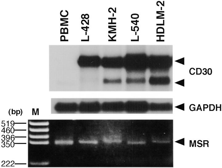
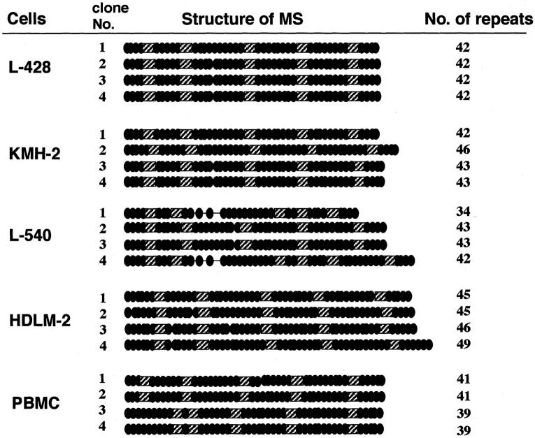
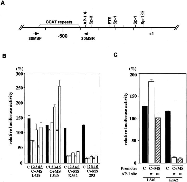
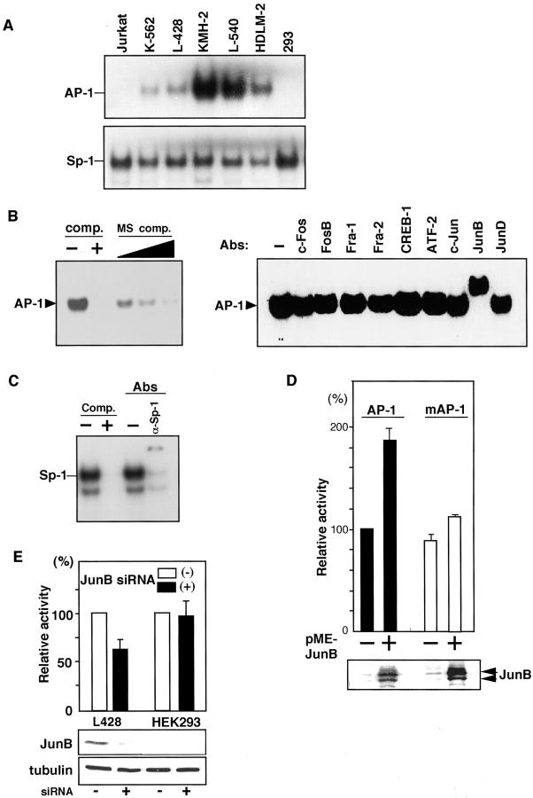
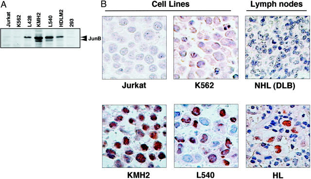
Overexpression of CD30 is the hallmark of Hodgkin and Reed-Sternberg (H-RS) cells and drives constitutive nuclear factor-kappaB activation that is the molecular basis for the pathophysiology of Hodgkin's lymphoma. Transcription of the CD30 gene is controlled by the core promoter that is driven by Sp-1 and the microsatellite sequences (MSs) that represses core promoter activity. To understand the mechanism(s) of CD30 overexpression in H-RS cells, we structurally and functionally characterized the CD30 MSs. Although the CD30 MS of H-RS cell lines was polymorphic, it was not truncated compared with that of control cells. A strong core promoter activity and constitutive Sp-1 binding were revealed in all cell lines examined irrespective of the levels of CD30 expression. In transient reporter gene assays, all MS clones derived from H-RS cell lines repressed the core promoter activity in unrelated cell lines, but not in the H-RS cell lines. An AP-1-binding site was found in the MS at nucleotide position of -377 to -371, the presence of which was found to relieve repression of the core promoter in H-RS cell lines but not in other tumor cell lines. H-RS cell lines showed constitutive and strong AP-1-binding activity, but other cell lines did not. The AP-1 complex contained JunB, whose overexpression activated reporter constructs driven by the CD30 promoter including the MSs, and was dependent on the AP-1 site. JunB expression was detected in H-RS cells in vitro and in vivo, but not in reactive cells or tumor cells of non-Hodgkin's lymphoma of diffuse large B-cell type. Transduction of JunB small interfering RNAs suppressed CD30 promoter activity in L428 cells but not in control cells. Taken together, overexpression and binding of JunB to the AP-1 site appear to relieve the repression of the core promoter by the CD30 MS in H-RS cells, which provide one basis for the constitutive overexpression of CD30 in Hodgkin's lymphoma.


Figures

Figure 1.
Figure 1.
CD30 mRNA expression and the length of MSs. Poly(A)-selected RNA (2 μg/lane) was analyzed by Northern blotting using cDNA probes of CD30 (top) and GAPDH (middle). Arrowheads indicate positions for two sizes of CD30 transcripts and GAPDH. PCR-amplified fragments of the CD30 MS were analyzed by agarose gel electrophoresis and ethidium bromide staining (bottom). The position for the PCR products of the MS is indicated by an arrowhead on the right. DNA size markers (M) are shown on the left.
Figure 2.
Figure 2.
Schematic representation of the structure of the CD30 MS. Nucleotide sequences of more than four clones from each cell line or a PBMC sample were determined. The CCAT repeat in the MS is indicated by a filled circle and the 5′-CCACTTATGCAT-3′ and 5′-CCACCTATGCAT-3′ motifs are indicated by hatched squares. The number of the CCAT repeats in each clone is indicated on the right.
Figure 3.
Figure 3.
Regulation of the CD30 promoter by the MS. A: Schematic representation of the CD30 promoter. Binding sites for transcription factors are indicated above the line. An asterisk indicates the position of the AP-1 site in the MS. Positions of the primers used to amplify the MS are shown below the line. B: Effects of the CD30 MS on the core promoter activity in various cell lines. Luciferase reporter constructs driven by the CD30 core promoter (−295 to + 198), with or without the CD30 MS, were transiently transfected with Renilla luciferase vector (pRL-TK). The promoter activities among different cell lines were compared with the results of parallel experiments with a firefly luciferase construct driven by the SV40 promoter and enhancer (pSV-Luc) and pRL-TK. The relative luciferase activities are expressed as percentages of those of pSV-Luc. C, a construct driven by the core promoter from PBMCs; C+MS, a construct driven by the core promoter with CD30 MS. The fragments used are as follows: clone number 1 of L428, number 2 of KMH-2, number 2 of L540, number 2 of HDLM-2, and number 1 of the control PBMCs in Figure 2 ▶ . The numbers 1 to 5 indicate luciferase constructs containing a CD30 core promoter with MS fragment derived from L428, KMH-2, L540, HDLM-2, and the control PBMCs, respectively. C: Effects of a mutation in the AP-1 site on the core promoter activities. Luciferase constructs having the CD30 core promoter alone (C) or with an MS fragment with a mutation in the AP-1 (C+MS, m) or without a mutation in the AP-1 (C+MS, w) were transfected into a H-RS cell line L540 and an unrelated cell line, K562. Luciferase activities were standardized using an SV40-driven luciferase construct as described above and expressed as a percentage. C, a core promoter-driven luciferase construct; C+MS, a construct driven by the core promoter with CD30 MS; w, the MS without a mutation; m, the MS with a mutation.
Figure 4.
Figure 4.
AP-1 activities in H-RS cells. A: AP-1- and Sp-1-binding activities. Nuclear extracts of H-RS cell lines and unrelated ones were studied by EMSA. Probes used are indicated on the left. B: Competition and supershift analyses of AP-1. Competition assays were done using a 50-fold molar excess of the unlabeled consensus Ap-1 probe or the unlabeled oligomer containing the AP-1 site in the CD30 MS at 50- to 150-fold molar excess (left). Comp., the consensus AP-1 oligomer; MS comp., oligomer containing the AP-1 site in the CD30 MS. AP-1 supershift assays were done using L-540 nuclear extracts (right). Antibodies used are indicated above the lanes. C: Competition and supershift analyses of Sp-1-binding complex using L540 nuclear extract. Comp., competition assay using 50-fold molar excess of the unlabeled consensus Sp-1 oligomer; Abs, supershift assay with anti-Sp-1 antibody. D: Responses to transduced JunB. Luciferase constructs driven by the CD30 promoter with or without a mutation in AP-1 site (pGL-CD30cMS and pGL-CD30cMSmA, respectively) was co-transfected with a JunB expression plasmid (pME-JunB) into K562 cells. pRL-TK was co-transfected to standardize transfection efficiency. AP-1, pGL-CD30cMS; mAP-1, pGL-CD30cMSmA. E: Effects of siRNA-mediated repression of endogenous JunB in H-RS cells on CD30 promoter activity. Twenty pmol of siRNA for JunB, 0.5 μg of reporter construct containing CD30 promoter (pGL-CD30cMS), and 0.2 μg of pRL-TK vector were transfected with 2.5 μl of Lipofectamine (Invitrogen). CD30 promoter activities were measured by Dual Luciferase assay kit (Promega) after 24 hours. Bottom, immunoblot analysis of JunB and tubulin expression.
Figure 5.
Figure 5.
Constitutive expression of JunB in H-RS cells in vitro and in vivo. A: Immunoblot analysis of Jun B. Whole cell lysates of four H-RS cell lines and three unrelated cell lines were examined. Positions of JunB are indicated on the right. B: Immunohistochemistry of JunB. Results of H-RS cell lines and control ones, and representative results of biopsied samples of classical HL and diffuse large B-cell NHL.

Similar articles

Cited by

References

    1. Gruss HJ, Kadin ME: Pathophysiology of Hodgkin’s disease: functional and molecular aspects. Baillieres Clin Haematol 1996, 9:417-446 - PubMed
    1. Cossman J, Annunziata CM, Barash S, Staudt L, Dillon P, He WW, Ricciardi-Castagnoli P, Rosen CA, Carter KC: Reed-Sternberg cell genome expression supports a B-cell lineage. Blood 1999, 94:411-416 - PubMed
    1. Kanzler H, Kuppers R, Hansmann ML, Rajewsky K: Hodgkin and Reed-Sternberg cells in Hodgkin’s disease represent the outgrowth of a dominant tumor clone derived from (crippled) germinal center B cells. J Exp Med 1996, 184:1495-1505 - PMC - PubMed
    1. Kuppers R, Rajewsky K: The origin of Hodgkin and Reed/Sternberg cells in Hodgkin’s disease. Annu Rev Immunol 1998, 16:471-493 - PubMed
    1. Kuppers R, Rajewsky K, Zhao M, Simons G, Laumann R, Fischer R, Hansmann ML: Hodgkin disease: Hodgkin and Reed-Sternberg cells picked from histological sections show clonal immunoglobulin gene rearrangements and appear to be derived from B cells at various stages of development. Proc Natl Acad Sci USA 1994, 91:10962-10966 - PMC - PubMed

Publication types