. 2003 Aug;163(2):643-52.
doi: 10.1016/S0002-9440(10)63691-7.

Neuron-specific activation of murine cytomegalovirus early gene e1 promoter in transgenic mice

Affiliations

Neuron-specific activation of murine cytomegalovirus early gene e1 promoter in transgenic mice

Yoshifumi Arai et al. Am J Pathol. 2003 Aug.

Abstract

The brain is the main target in congenital cytomegalovirus (CMV) infection and immunocompromised patients. No definite evidence that a CMV has special affinity for the central nervous system (CNS) has been published. Here, we generated transgenic mice with an e1 promoter/enhancer region connected to the reporter gene lacZ. Surprisingly, expression of the transgene was completely restricted to the CNS in all lines of transgenic mice. The transgene was expressed in subpopulation of neurons in the cerebral cortex, hippocampus, diencephalon, brainstem, cerebellum, and spinal cord in all of the lines. Non-neuronal cells in the CNS were negative for transgene expression. Activation of the transgene was first observed in neurons of mesencephalon in late gestation, and then the number of positive neurons increased in various parts of the brain as development proceeded. Upon infection of the transgenic mouse brains with MCMV, the location of the activated neurons became more extensive, and the number of such neurons increased. These results suggest that there are host factor(s) that directly activate the MCMV early gene promoter in neurons. This neuron-specific activation may be associated with persistent infection in the brain and may be responsible for the neuronal dysfunction and neuronal cell loss caused by CMV infection.


Figures

Figure 1.
Figure 1.
Structure of the transgene, and Southern blot analysis. A: Construct used to generate MCMV-e1-pro-lacZ transgenic lines (transgene; 5.2 kb). The MCMV e1 (M112–113) promoter (MCMV-e1-pro) (nucleotides − 1534 to + 38) was inserted into the pnlacF vector, containing nuclear localization signal (N) and polyadenylation signals from the mouse protamine (mP1) gene. For the preparation of transgene, the vector was digested with KpnI and BglII. B: Southern blotting of the tail DNA from the four transgenic lines. Aliquots of 10 μg of tail DNA were digested with BamHI, electrophoresed in a 0.9% agarase gel, transferred to a nylon membranes, and probed with a 33P-labeled BamHI fragment (3.0 kb; A). Control lanes show increasing amounts: 0 copy (lane 1), 1 copy (lane 2), 5 copies (lane 3), 10 copies (lane 4), 20 copies (lane 5) of BamHI-digested mixtures of MCMV-e1-pro-lacZ with 10 μg of normal mouse tail DNA. Lane 6, Tg-029; lane 7, Tg-041; lane 8, Tg046; lane 9, Tg-033.
Figure 2.
Figure 2.
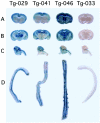
β-galactosidase expression in the CNS in 4-week-old transgenic lines. The sliced brains and spinal cords from three transgenic lines (Tg-029, Tg-041, Tg-046, and Tg-033) were stained for X-Gal. A: Coronal cerebral slices through frontal lobe. B: Coronal cerebral slices through the mammillary bodies. C: Sagittal slices of the brainstem and cerebellum. D: Spinal cords. In Tg-033, expression of the transgene was not detected in normal conditions.
Figure 3.
Figure 3.
Expression of β-galactosidase of the organs from Tg-029 assayed quantitatively by the LightCycler RT-PCR. A: RNAs extracted from the various organs of Tg-029 mice, were reverse-transcribed into cDNA, then assayed by the LightCycler using the primers specific for β-gal and for HPRT. Copy numbers were calculated with reference to standard sample. The copy numbers of β-gal cDNA from organs were expressed as divided by those of mouse HPRT cDNA for normalization. Data were averages of three experiments. B: The same cDNAs were subjected to ordinary PCR using same primers. The PCR products were electrophoresed on 2% agarose gels.
Figure 4.
Figure 4.
Immunohistochemical staining of expression of MCMV-e1-pro-lacZ in transgenic mice. The CNS tissues from a 4-week-old transgenic Tg-029 line were fixed in 4% PFA and embedded in paraffin. Deparaffinized sections were incubated with the anti-β-galactosidase antibody (anti-β-gal), followed by horseradish peroxidase (HRP)-conjugated goat anti-rabbit IgG, and then colored with 3-amino-9-ethyl carbazole (AEC). A and B: Cerebral cortex. C and D: Hippocampus dentate gyrus (DG). E and F: Diencephalon. G and H: Cerebellar cortex. Purkinje cells (arrows). I: Brainstem. J: Spinal cord. Bars, 100 μm (A, C, E, G, I, and J); 30 μm (B, D, F, and H).
Figure 5.
Figure 5.
Immunohistochemical double staining of β-galactosidase and neural markers. To confirm that β-gal-positive cells are neuronal cells, the cerebral cortex of a 4-week-old Tg-029 mouse was double-stained using anti-β-gal Ab and anti-neuron-specific enolase (NSE) Ab (A), anti-β-tubulin (B), anti-MAP2 (C), or anti-GFAP Ab (D). The brain that had been incubated with anti-β-gal Ab was colored with fast blue BB (blue), then incubated with antibodies specific to neural markers and colored with AEC (red). Bars, 30 μm.
Figure 6.
Figure 6.
Alternation of activation of the MCMV-e1-promoter during the brain development. Expression of the transgene was examined immunohistochemically using anti-β-gal Ab in Tg-029 line at E18.5 (A and E), P3 (B and F), P7 (C and G), and P28 (D and H). In the cerebral cortex (A–D) and hippocampus (E–H), positive cells gradually increase and the distribution expanded as the growth proceeded. Bars, 100 μm (A–D), 200 μm (E–H).
Figure 7.
Figure 7.
Induction of activation of the MCMV-e1-promoter in the Tg-033 brain by MCMV infection. Intracerebral injection of the transgenic mice with MCMV (105 PFU) was performed within 24 hours after their birth, and the brains were examined at P7. Intracerebral injection of the same litter of the transgenic mice with MEM was also performed in the same manner for uninfected control. A–D: Cerebral cortex. E–H: Hippocampus. The brain sections were immunohistochemically stained using anti-β-gal Ab, colored with AEC (red) (A and E, uninfected; B and F, infected; arrows, β-gal-positive cells). C and G: Adjacent section of B and F were stained with mAb D5, specific to the MCMV early antigen, colored with fast blue BB (blue). D and H: The β-gal-positive cells induced by MCMV infection were (red) were double-stained with the rat mAb D5 (blue). Arrows, double-positive cells. Arrowheads, β-gal-negative-infected cells. *, cells singly positive for β-gal. Bars, 100 μm (A–C and E–G); D and H, 30 μm.
Figure 8.
Figure 8.
Quantitative comparison of β-gal-expressed neurons between uninfected and MCMV-infected cerebral cortex and hippocampus in the transgenic lines (Tg-029, Tg-046, and Tg-033). Transgenic mouse brains from same littermate were injected with MEM (A) and with MCMV (10 PFU) (B) in the same manner as Figure 7 ▶ . C: β-gal-expressed neurons double-stained with mAb D5, specific for MCMV early nuclear antigen. D: Virus-infected cells stained with mAb D5. Positive cell numbers were expressed per cortex area or hippocampus of each cerebral hemisphere. The data of three animals in each transgenic line were averaged. *, P < 0.05 (Student’s t-test).

Similar articles

Cited by

References

    1. Weller TH: The cytomegalovirus: ubiquitous agents with protean clinical manifestations. N Engl J Med 1971, 285:203-214 - PubMed
    1. Becroft DMO: Prenatal cytomegalovirus infection: epidemiology, pathology, and pathogenesis. Perspect Pediatr Pathol 1981, 6:203-241 - PubMed
    1. Stagno S, Pass RF, Cloud G, Bright WJ, Henderson RE, Walton PD, Veren DA, Page F, Alford CA: Primary cytomegalovirus infection in pregnancy: incidence, transmission to fetus, and clinical outocome. JAMA 1986, 256:1904-1908 - PubMed
    1. Ho M: Congenital and perinatal human cytomegalovirus infection. Ho M eds. Cytomegalovirus: Biology and Infection. 1991:pp 205-227 Plenum New York
    1. Pass RF, Stagno S, Myers GJ, Alford CA: Outcome of symptomatic congenital cytomegalovirus infection: results of long-term longitudinal follow-up. Pediatrics 1980, 66:758-762 - PubMed

Publication types

LinkOut - more resources