. 2003 Aug;163(2):683-90.
doi: 10.1016/S0002-9440(10)63695-4.

Characterization of methylthioadenosin phosphorylase (MTAP) expression in malignant melanoma

Affiliations

Characterization of methylthioadenosin phosphorylase (MTAP) expression in malignant melanoma

Iris Behrmann et al. Am J Pathol. 2003 Aug.

Abstract

Homozygous deletions of human chromosomal region 9p21 occur frequently in malignant melanoma and are associated with the loss of the tumor suppressor genes p16(INK4a) and p15(INK4b). In the same chromosomal region the methylthioadenosine phosphorylase (MTAP) gene is localized and therefore may also serve as a tumor suppressor gene. The aim of this study was to analyze MTAP mutations and expression patterns in malignant melanomas. To examine the MTAP gene and expression of MTAP protein we screened 9 human melanoma cell lines and primary human melanocytes by reverse transcriptase-polymerase chain reaction, sequencing, and immunoblotting. Analyzing the melanoma cell lines we found significant down-regulation of MTAP mRNA expression. In only one cell line, HTZ19d, this was due to homozygous deletion of exon 2 to 8 whereas in the other cell lines promoter hypermethylation was detected. MTAP expression was further analyzed in vivo by immunohistochemical staining of 38 tissue samples of benign melanocytic nevi, melanomas, and melanoma metastases. In summary, we demonstrate significant inverse correlation between MTAP protein expression and progression of melanocytic tumors as the amount of MTAP protein staining decreases from benign melanocytic nevi to metastatic melanomas. Our results suggest an important role of MTAP inactivation in the development of melanomas. This finding may be of great clinical significance because recently an association between MTAP activity and interferon sensitivity has been suggested.


Figures

Figure 1.
Figure 1.
Amplification of the complete coding region of MTAP mRNA by PCR. The complete coding region was specifically amplified by RT-PCR. All melanoma cells except of HTZ19d were shown to express full-length MTAP mRNA. Strength of expression cannot be estimated by this experiment as the PCR was carried out to saturation to illustrate full-length mRNA expression and to obtain enough product for sequencing.
Figure 2.
Figure 2.
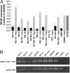
MTAP expression in melanoma cell lines compared to primary melanocytes. A: By real-time PCR the amount of MTAP mRNA expression was carefully quantified. All melanoma cell lines showed significant reduction in MTAP expression compared to human primary melanocytes set as 1 (black bars). In HTZ19d this was due to loss of the genomic region. By treatment with the demethylating agent 5-azacytidine, re-expression of MTAP was found in all melanoma cell lines except of HTZ19d (gray bars). B: By PCR amplification the methylation status of the MTAP promoter was analyzed. Genomic DNA was incubated with NotI which cuts in the CpG island of the unmethylated MTAP promoter. Methylation interferes with NotI activity (gDNA->NotI->PCR). As a control for integrity of the genomic DNA, DNA without NotI digestion was analyzed (gDNA -> PCR). PCR amplification was successfully obtained with genomic DNA of all melanoma cell lines after NotI digestion but not in normal human epidermal keratinocytes (NHEK). Keratinocytes were used as control for strongly MTAP-expressing cells (see Figure 4 ▶ , immunohistochemistry).
Figure 3.
Figure 3.
Western blot analysis of MTAP protein expression in melanoma cell lines. Expression of MTAP protein was detected in primary human melanocytes. In melanoma cell lines the expression was reduced. As loading control the blot was counterstained with a β-actin antibody.
Figure 4.
Figure 4.
Immunostaining of MTAP in human melanocytic tumors. A and B: Strong cytoplasmatic immunosignals in benign melanocytic nevi (blue arrows). C and D: Focally positive-(blue arrows) and negative-staining results (black arrow in D) in primary cutaneous melanomas. E and F: Negative or weak staining pattern in metastases from cutaneous melanomas. Completely unstained melanoma cells in F are marked by black arrows. Small round cells underneath the metastatic infiltrate represent resident lymphocytes of the lymph node. Immunosignals were developed in red (AEC) and are distinct from the brown melanin pigment. Slides were counterstained with hemalaun and magnified ×250.
Figure 5.
Figure 5.
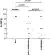
Evaluation of MTAP staining. Analogous to the Remmele score (see Material and Methods) intensity and percentage of stained cells was evaluated. The maximal score is 12 for strong staining of more than 80% stained cells. 9 nevi, 15 primary melanomas, and 14 melanoma metastases were evaluated.
Figure 6.
Figure 6.
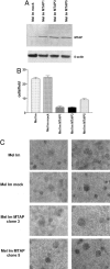
Functional relevance of MTAP expression in malignant melanoma. A: Western blot analysis of the Mel Im cell clones stably transfected with an MTAP expression plasmid. All cell clones (Mel Im MTAP 1, 3 and 5) showed expression of MTAP whereas MTAP expression in the mock transfected cell clone (Mel Im mock) stayed unchanged. B: Analysis of the cell clones re-expressing MTAP revealed strong reduction of their invasive and migratory potential as shown by Boyden Chamber assays. C: Colony forming assays revealed no differences in the ability of the cell clones to form soft agar colonies.

References

    1. Mowen KA, Tang J, Zhu W, Schurter BT, Shuai K, Herschman HR, David M: Arginine methylation of STAT1 modulates IFNα/β-induced transcription. Cell 2001, 104:731-741 - PubMed
    1. Christopher SA, Diegelman P, Porter CW, Kruger WD: Methylthioadenosine phosphorylase, a gene frequently codeleted with p16(cdkN2a/ARF), acts as a tumor suppressor in a breast cancer cell line. Cancer Res 2002, 62:6639-6644 - PubMed
    1. Garcia-Castellano JM, Villanueva A, Healey JH, Sowers R, Cordon-Cardo C, Huvos A, Bertino JR, Meyers P, Gorlick R: Methylthioadenosine phosphorylase gene deletions are common in osteosarcoma. Clin Cancer Res 2002, 8:782-787 - PubMed
    1. Wong YF, Chung TK, Cheung TH, Nobori T, Chang AM: MTAP gene deletion in endometrial cancer. Gynecol Obstet Invest 1998, 45:272-276 - PubMed
    1. Hori Y, Hori H, Yamada Y, Carrera CJ, Tomonaga M, Kamihira S, Carson DA, Nobori T: The methylthioadenosine phosphorylase gene is frequently co-deleted with the p16INK4a gene in acute type adult T-cell leukemia. Int J Cancer 1998, 75:51-56 - PubMed

Publication types

Substances