. 2003 Jul 21;162(2):211-22.
doi: 10.1083/jcb.200212004.

Vacuole membrane fusion: V0 functions after trans-SNARE pairing and is coupled to the Ca2+-releasing channel

Affiliations

Vacuole membrane fusion: V0 functions after trans-SNARE pairing and is coupled to the Ca2+-releasing channel

Martin J Bayer et al. J Cell Biol. .

Abstract

Pore models of membrane fusion postulate that cylinders of integral membrane proteins can initiate a fusion pore after conformational rearrangement of pore subunits. In the fusion of yeast vacuoles, V-ATPase V0 sectors, which contain a central cylinder of membrane integral proteolipid subunits, associate to form a transcomplex that might resemble an intermediate postulated in some pore models. We tested the role of V0 sectors in vacuole fusion. V0 functions in fusion and proton translocation could be experimentally separated via the differential effects of mutations and inhibitory antibodies. Inactivation of the V0 subunit Vph1p blocked fusion in the terminal reaction stage that is independent of a proton gradient. Deltavph1 mutants were capable of docking and trans-SNARE pairing and of subsequent release of lumenal Ca2+, but they did not fuse. The Ca2+-releasing channel appears to be tightly coupled to V0 because inactivation of Vph1p by antibodies blocked Ca2+ release. Vph1 deletion on only one fusion partner sufficed to severely reduce fusion activity. The functional requirement for Vph1p correlates to V0 transcomplex formation in that both occur after docking and Ca2+ release. These observations establish V0 as a crucial factor in vacuole fusion acting downstream of trans-SNARE pairing.


Figures

Figure 1.
Figure 1.
Vacuolar structure in V-ATPase mutants. (A) Yeast cells (BY4742) containing the indicated deletions of different V-ATPase subunits were grown logarithmically in liquid YPD medium, stained with FM4–64, and analyzed by fluorescence microscopy as described in Materials and methods. (B) Quantitation of vacuole morphology. The number of vacuolar vesicles (stained by FM4–64) per cell was determined for the strains shown in A. For each experiment, 200 cells per strain were analyzed and grouped into the indicated categories. Two experiments were averaged. Bar, 5 μm.
Figure 1.
Figure 1.
Vacuolar structure in V-ATPase mutants. (A) Yeast cells (BY4742) containing the indicated deletions of different V-ATPase subunits were grown logarithmically in liquid YPD medium, stained with FM4–64, and analyzed by fluorescence microscopy as described in Materials and methods. (B) Quantitation of vacuole morphology. The number of vacuolar vesicles (stained by FM4–64) per cell was determined for the strains shown in A. For each experiment, 200 cells per strain were analyzed and grouped into the indicated categories. Two experiments were averaged. Bar, 5 μm.
Figure 2.
Figure 2.
Fusion activity of vacuoles from strains deficient in V1 sector (Δvma1) or in V0 sector (Δvph1). (A) Vacuoles from the indicated mutants (DKY 6281 background) were fused with wild-type vacuoles (BJ3505). Incubations were performed in the presence or absence of untreated cytosol (C) or of cytosol immunodepleted for Vma1p and Vma2p (dC). Fusion was assayed after 70 min at 27°C. The fusion activities of control reactions (asterisk) were set to 100%. The activity of the samples on ice was set to 0% (n = 5). Fusion activities of the wild-type–wild-type combination ranged from 2.2 to 4.1 U. (B) Western blot of equal protein amounts of cytosol (C) and immunodepleted cytosol (dC) against Vma1p, Vma2p, and the cytosolic marker protein phosphoglycerate kinase (PGK), verifying the absence of V1 sectors in the depleted cytosol.
Figure 2.
Figure 2.
Fusion activity of vacuoles from strains deficient in V1 sector (Δvma1) or in V0 sector (Δvph1). (A) Vacuoles from the indicated mutants (DKY 6281 background) were fused with wild-type vacuoles (BJ3505). Incubations were performed in the presence or absence of untreated cytosol (C) or of cytosol immunodepleted for Vma1p and Vma2p (dC). Fusion was assayed after 70 min at 27°C. The fusion activities of control reactions (asterisk) were set to 100%. The activity of the samples on ice was set to 0% (n = 5). Fusion activities of the wild-type–wild-type combination ranged from 2.2 to 4.1 U. (B) Western blot of equal protein amounts of cytosol (C) and immunodepleted cytosol (dC) against Vma1p, Vma2p, and the cytosolic marker protein phosphoglycerate kinase (PGK), verifying the absence of V1 sectors in the depleted cytosol.
Figure 3.
Figure 3.
Detection of V-ATPase–independent proton uptake activity by acridine orange. (A) Concentration dependence of the assay. Proton uptake activity of the vacuoles was assayed by measuring acridine orange absorption. Fusion reactions were started in the presence of 15 μM acridine orange and different vacuole concentrations to vary the dye to vacuole ratio. Where indicated (start), the ATP regenerating system and concanamycin A had been added. At the end of the assay period, FCCP (30 μM) was added to dissipate the proton gradient. (B) Assay of mutant vacuoles. Vacuoles from wild-type, Δvph1, and Δvma2 cells were assayed for proton uptake activity as in A. 54 pmol acridine orange (final concentration 15 μM) were used per μg vacuoles.
Figure 4.
Figure 4.
Fusion and apparent proton translocation activity of vacuoles from vph1 deletion strains (Δvph1). (A) Fusion and apparent proton translocation activity (B) were examined in parallel fusion reactions using wild-type or Δvph1 vacuoles in the presence or absence of concanamycin A (Conc.A; 1 μM) or FCCP (30 μM). Fusion and pump activities of wild-type vacuoles (asterisk) were set to 100% (n = 3). Fusion activities of these control samples ranged from 2.5 to 3.8 U.
Figure 5.
Figure 5.
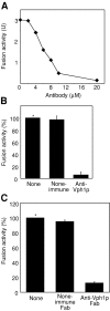
Inhibition by antibodies to Vph1p. (A) Different concentrations of affinity-purified antibodies to Vph1p (from goat) were added to a standard fusion reaction without cytosol. The reaction was started after preincubation for 10 min on ice by adding ATP. Fusion was assayed after 70 min at 27°C. (B) Nonimmune antibodies do not interfere with vacuole fusion. Standard fusion reactions were run with 20 μM IgG purified from nonimmune or anti-Vph1p sera from goats, or with control buffer only, and assayed after 70 min at 27°C (n = 6). (C) Inhibition of fusion by Fab fragments of antibodies to Vph1p. Standard fusion reactions were incubated with Fab fragments derived either from nonimmune antibodies or from antibodies to Vph1p. Fusion was assayed as in the legend to Fig. 2 (n = 3). Fusion activities of the control samples (asterisk) ranged from 3.2 to 5.1 U, and those of ice values ranged from 0.2 to 0.3 U. Inhibitor concentrations were 45 μM anti-Vph1p Fab and 45 μM nonimmune Fab.
Figure 6.
Figure 6.
Influence of antibodies to Vph1p on the apparent proton translocation and fusion activity of intact vacuoles. Standard fusion reactions were run in the presence or absence of the indicated inhibitors. Pump activity was determined with the BCECF assay as described in Materials and methods. Fusion activities were assayed in parallel samples as in Fig. 2 (n = 4). Fusion activities of control samples ranged from 3.3 to 4.8 U. Inhibitors were FCCP (30 μM), anti-Vph1p (20 μM), and nonimmune antibodies (20 μM).
Figure 7.
Figure 7.
Fusion of vacuoles without V1 sector is inhibited by anti-Vph1p. (A) Vacuoles were extracted with SCN/ATP to remove V1. Fusion activity was assayed in the absence or presence of the indicated inhibitors as in the legend to Fig. 2 (n = 3). Fusion activities of the control samples (asterisk) ranged from 0.75 to 1.4 U. (B) Apparent proton pumping activity of untreated or stripped vacuoles was determined as described in Materials and methods. The activity of untreated vacuoles was set to 100%. Inhibitors were GTPγS (2 mM), anti-Vph1p (20 μM), nonimmune antibodies (20 μM), and anti-Sec18p (2 μM).
Figure 8.
Figure 8.
Kinetic resolution of Vph1p requirement for fusion. (A) Standard fusion reactions without cytosol were started at 27°C. At the indicated times, inhibitors or control buffer were added. The samples were left on ice for 10 min. Then, they were transferred to 27°C or left on ice for the remainder of the 70-min reaction period. After 70 min, fusion activity was assayed. (B) Trans-SNARE pairing. Fusion reactions with vacuoles from strains SBY521 (Δvam3 Δvph1) or no. 418 (Δvam3 VPH1) and no. 120 (Δnyv1 VPH1) were incubated (50 min, 27°C) with the indicated inhibitors at 27°C or left on ice. Then, the membranes were reisolated, solubilized, and assayed for trans-SNARE complexes by determining the amounts of the v-SNARE Nyv1p coimmunoprecipitating with the t-SNARE Vam3p. (C) Standard fusion reactions without cytosol were incubated at 27°C in the presence of 1 mM MgCl2 and 5 mM BAPTA for 30 min. The reactions were chilled on ice, and vacuoles were reisolated (10,000 g, 2 min, 2°C). Vacuoles were resuspended in fusion buffer with cytosol and 200 μM CaCl2 but without ATP. Aliquots were preincubated for 5 min on ice with the indicated inhibitors and incubated further for 70 min at 27°C. Fusion activities were assayed and plotted as in the legend to Fig. 2 (n = 3). Activities of the control sample ranged from 1.1 to 1.9 U. Inhibitors were BAPTA (5 mM), GTPγS (2 mM), anti-Vam3p (2 μM), Gdi1p (5 μM), anti-Vph1p (20 μM), and FCCP (30 μM).
Figure 8.
Figure 8.
Kinetic resolution of Vph1p requirement for fusion. (A) Standard fusion reactions without cytosol were started at 27°C. At the indicated times, inhibitors or control buffer were added. The samples were left on ice for 10 min. Then, they were transferred to 27°C or left on ice for the remainder of the 70-min reaction period. After 70 min, fusion activity was assayed. (B) Trans-SNARE pairing. Fusion reactions with vacuoles from strains SBY521 (Δvam3 Δvph1) or no. 418 (Δvam3 VPH1) and no. 120 (Δnyv1 VPH1) were incubated (50 min, 27°C) with the indicated inhibitors at 27°C or left on ice. Then, the membranes were reisolated, solubilized, and assayed for trans-SNARE complexes by determining the amounts of the v-SNARE Nyv1p coimmunoprecipitating with the t-SNARE Vam3p. (C) Standard fusion reactions without cytosol were incubated at 27°C in the presence of 1 mM MgCl2 and 5 mM BAPTA for 30 min. The reactions were chilled on ice, and vacuoles were reisolated (10,000 g, 2 min, 2°C). Vacuoles were resuspended in fusion buffer with cytosol and 200 μM CaCl2 but without ATP. Aliquots were preincubated for 5 min on ice with the indicated inhibitors and incubated further for 70 min at 27°C. Fusion activities were assayed and plotted as in the legend to Fig. 2 (n = 3). Activities of the control sample ranged from 1.1 to 1.9 U. Inhibitors were BAPTA (5 mM), GTPγS (2 mM), anti-Vam3p (2 μM), Gdi1p (5 μM), anti-Vph1p (20 μM), and FCCP (30 μM).
Figure 9.
Figure 9.
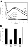
Vacuolar Ca2 + release during fusion. (A) Inhibition by antibodies to Vph1p. Standard fusion reactions were started, and Ca2+ was monitored continuously as described in the Materials and methods. Before the reactions were started, vacuoles had been preincubated with one of the indicated inhibitors, or with one of the following buffers only (3 min, 0°C): anti-Sec18p (2 μM), anti-Sec17p (2 μM), anti-Vph1p (20 μM), and nonimmune antibodies (20 μM). (B) Effect of vph1 deletion. Vacuoles were prepared from wild-type and Δvph1 cells and incubated under fusion conditions in the presence or absence of 5 μM Gdi1p. Ca2+ efflux was monitored as in A, and peak signals were plotted.

References

    1. Adachi, I., K. Puopolo, N. Marquez-Sterling, H. Arai, and M. Forgac. 1990. Dissociation, cross-linking, and glycosylation of the coated vesicle proton pump. J. Biol. Chem. 265:967–973. - PubMed
    1. Adamo, J.E., G. Rossi, and P. Brennwald. 1999. The Rho GTPase Rho3 has a direct role in exocytosis that is distinct from its role in actin polarity. Mol. Biol. Cell. 10:4121–4133. - PMC - PubMed
    1. Adamo, J.E., J.J. Moskow, A.S. Gladfelter, D. Viterbo, D.J. Lew, and P.J. Brennwald. 2001. Yeast Cdc42 functions at a late step in exocytosis, specifically during polarized growth of the emerging bud. J. Cell Biol. 155:581–592. - PMC - PubMed
    1. Adler, E.M., G.J. Augustine, S.N. Duffy, and M.P. Charlton. 1991. Alien intracellular calcium chelators attenuate neurotransmitter release at the squid giant synapse. J. Neurosci. 11:1496–1507. - PMC - PubMed
    1. Arai, H., S. Pink, and M. Forgac. 1989. Interaction of anions and ATP with the coated vesicle proton pump. Biochemistry. 28:3075–3082. - PubMed

Publication types

MeSH terms