. 2003 Jul 21;162(2):281-91.
doi: 10.1083/jcb.200212141.

PAK1 phosphorylation of MEK1 regulates fibronectin-stimulated MAPK activation

Affiliations

PAK1 phosphorylation of MEK1 regulates fibronectin-stimulated MAPK activation

Jill K Slack-Davis et al. J Cell Biol. .

Abstract

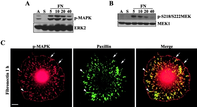
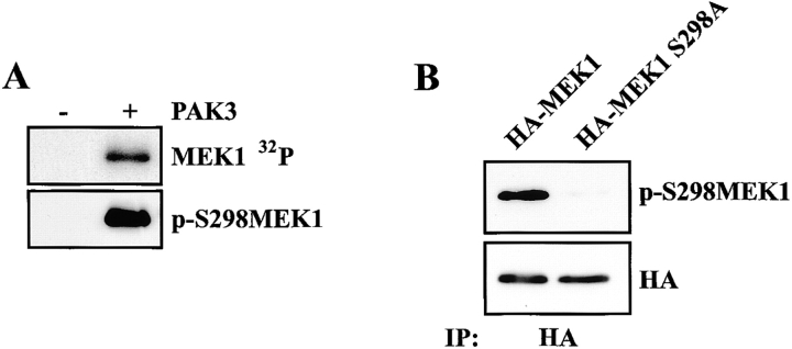
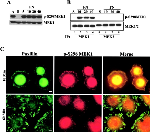
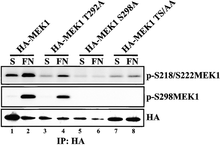
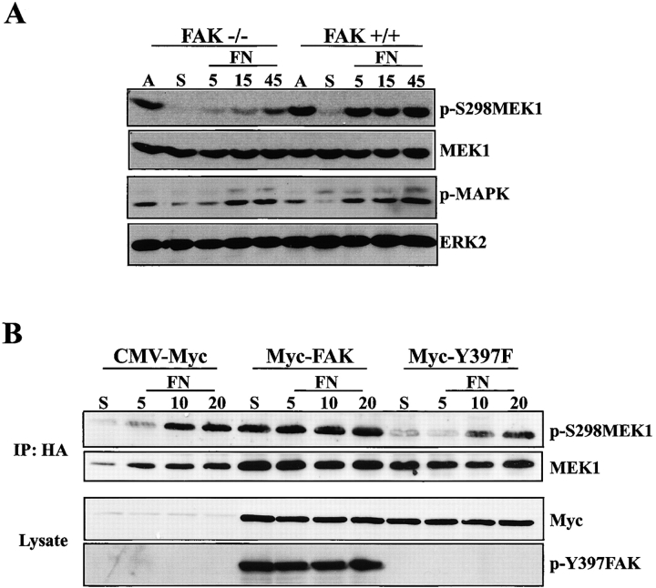
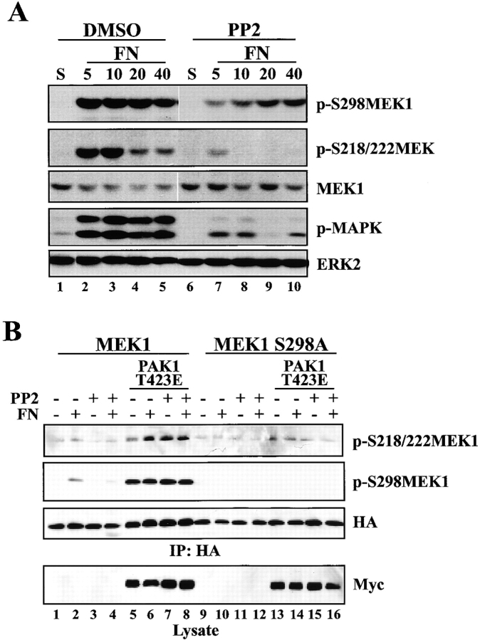
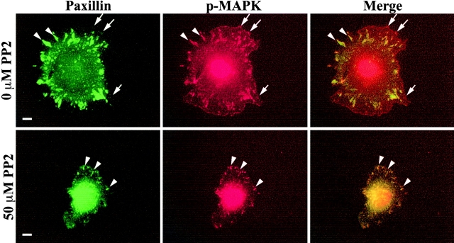
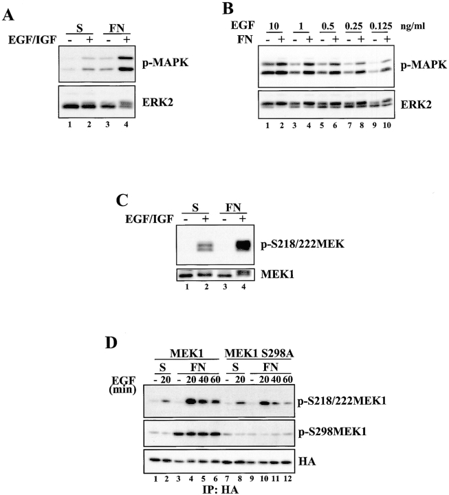
Activation of the Ras-MAPK signal transduction pathway is necessary for biological responses both to growth factors and ECM. Here, we provide evidence that phosphorylation of S298 of MAPK kinase 1 (MEK1) by p21-activated kinase (PAK) is a site of convergence for integrin and growth factor signaling. We find that adhesion to fibronectin induces PAK1-dependent phosphorylation of MEK1 on S298 and that this phosphorylation is necessary for efficient activation of MEK1 and subsequent MAPK activation. The rapid and efficient activation of MEK and phosphorylation on S298 induced by cell adhesion to fibronectin is influenced by FAK and Src signaling and is paralleled by localization of phospho-S298 MEK1 and phospho-MAPK staining in peripheral membrane-proximal adhesion structures. We propose that FAK/Src-dependent, PAK1-mediated phosphorylation of MEK1 on S298 is central to the organization and localization of active Raf-MEK1-MAPK signaling complexes, and that formation of such complexes contributes to the adhesion dependence of growth factor signaling to MAPK.


Figures

Figure 1.
Figure 1.
Adhesion stimulates MAPK and MEK phosphorylation. REF52 cells were either continuously adherent (A) or suspended (S) and plated on FN for 5, 10, 20, or 40 min. Whole cell lysates were blotted with antiserum specific for (A) phosphorylated MAPK (p-MAPK; top) or ERK2 (bottom), or (B) MEK1 phosphorylated on S218/S222 (p-S218/222MEK1; top) or MEK1 (bottom). (C) REF52 cells were suspended for 1 h and plated on FN for 1 h before co-staining for p-MAPK (red) and paxillin (green). The arrows indicate focal complex-like structures containing p-MAPK; arrowheads indicate paxillin-containing focal adhesions. Bar, 10 μm.
Figure 2.
Figure 2.
PAK directs MEK1 S298 phosphorylation in vitro. (A) In vitro kinase assays were performed using recombinant MEK1 and recombinant PAK3 as described in Materials and methods. (B) REF52 cells were transfected with HA-tagged wild-type MEK1 or HA-MEK1 S298A, suspended for 1 h and allowed to adhere to FN for 10 min. Anti-HA immunoprecipitates were analyzed and blotted with p-S298MEK1 (top) or MEK1 (bottom).
Figure 3.
Figure 3.
MEK1 S298 phosphorylation is regulated by cell adhesion. (A) REF52 cells were treated as described in Fig. 1. Whole cell lysates were blotted with antiserum specific for phospho-S298 MEK1 (p-S298MEK1; top) or MEK1 (bottom). (B) REF52 cells were suspended (S) and replated on FN for 10, 20, or 40 min. Anti-MEK1 or anti-MEK2 antiserum was used to immunoprecipitate endogenous proteins, which were subsequently blotted with anti– p-S298MEK1 or anti-MEK1/2. (C) REF52 cells were suspended and plated on FN for 10 min or 1 h before co-staining for p-S298MEK1 (red) and paxillin (green). The intense staining in the center of the cell represents perinuclear staining that was over-exposed to visualize peripheral structures. Bars, 10 μm.
Figure 4.
Figure 4.
PAK regulates adhesion-dependent MEK1 S298 phosphorylation. (A) COS-1 cells were transfected with myc-tagged kinase-defective PAK1 (Myc-PAK1-KR) or empty vector control together with HA-tagged MEK1. Cells were suspended (S) or plated on FN for 10, 20, or 30 min. Immunoprecipitates were formed using HA antiserum and blotted for p-S298MEK1 or MEK1. PAK1 expression was determined from whole cell lysates of transfected cells by immunoblotting with Myc antiserum. (B) REF52 cells were transiently cotransfected with HA-MEK1 together with myc-PAK1 T423E or the appropriate empty vector. Cells were suspended for 90 min (S) and allowed to adhere to FN-coated dishes for 20 min (FN). Anti-HA immunoprecipitates were formed and blotted with HA antiserum (middle), and subsequently with anti-pS298MEK1 (top). Western blotting of lysates with anti-myc antiserum confirmed expression of activated PAK1 (bottom).
Figure 5.
Figure 5.
Phosphorylation of MEK1 on S298 regulates MEK1 activation. REF52 cells were transiently transfected with HA-MEK1, HA-MEK1 T292A, HA-MEK1 S298A, or HA-MEK1 T292A/S298A. Cells were suspended for 90 min (S) and plated on FN for 20 min (FN). Anti-HA immunoprecipitates were formed and blotted with anti-p-S218/S222MEK1 (top), anti-pS298MEK1 (middle), or anti-HA antiserum (bottom).
Figure 6.
Figure 6.
FAK signaling regulates MEK1 S298 phosphorylation. (A) FAK-null fibroblasts or cells from wild-type littermate controls were suspended (S), plated on FN for 5, 15, or 45 min, or remained adherent (A). Whole cell lysates were blotted with p-S298MEK1, MEK1, p-MAPK, or ERK2 antisera. (B) FAK-null fibroblasts transfected with myc-tagged wild-type FAK, FAK Y397F, or empty vector control together with HA-tagged wild-type MEK1 were suspended (S) and replated on FN for 5, 10, or 20 min. Immunoprecipitates were formed using HA antiserum and blotted for p-S298MEK1 or MEK1. FAK expression was determined from whole cell lysates of transfected cells using Myc antiserum. FAK autophosphorylation was determined by immunoblotting with Y397 FAK phospho-specific antibodies.
Figure 7.
Figure 7.
Src inhibition alters PAK-mediated MEK phosphorylation. (A) REF52 cells were incubated in suspension with 50 μM PP2 or DMSO control (S) and plated on 10 μg/ml FN for 5, 10, 20, or 40 min in the continued presence or absence of PP2. Whole cell lysates were blotted with p-S298MEK1, p-S218/S222MEK1, MEK1, p-MAPK, or ERK2 antisera. Densitometric analysis demonstrated that the intensity of p-S298MEK1 after a 5-min plating on FN in the presence of 50 μM PP2 was ∼28% the level of the DMSO-treated control when normalized to an MEK1 loading control. The observed decrease in p-MAPK levels in lane 9 was not reproducible. (B) REF52 cells were cotransfected with HA-tagged MEK1 constructs and PAK1 T423E or vector control, incubated in suspension with 50 μM PP2 or DMSO control (S) and plated on FN for 20 min. Anti-HA immunoprecipitates were formed and blotted with pS218/S222 antiserum (top), HA antiserum (second [from top] panel), and subsequently with anti-pS298MEK1 (third [from top] panel). Western blotting of lysates with anti-myc antiserum confirmed expression of activated PAK1 (bottom).
Figure 8.
Figure 8.
Adhesion-dependent c-Raf and PAK activity are affected by inhibiting Src. (A) c-Raf activity was determined from REF52 cells incubated in suspension with 50 μM PP2 or DMSO solvent control (S) and plated on FN for 5 or 10 min. Immunoprecipitates were formed using either c-Raf antiserum, or matched control antiserum raised against the GAL4 transactivation domain, and kinase assays were performed with kinase-defective GST-MEK1 as described in Materials and methods (top). A reaction blank (lane B) confirms the absence of MEK kinase activity in the substrate and c-Raf antiserum, and a reaction lacking GST-MEK1 (lane 8) confirms that MEK1 is the phosphorylated substrate in these assays. Loading controls (bottom) demonstrate similar levels of c-Raf protein in the appropriate assays. (B) PAK1 activity was determined as described above for c-Raf activity (A) except cells were also plated for 5, 10, or 20 min and immunoprecipitates were formed using PAK1 antiserum. PAK1 activity was assessed by blotting the recombinant MEK1 substrate with the p-S298 antiserum (top). The PAK1 loading control is shown in the bottom panel. Lane B denotes a reaction blank in which PAK1 antibody was incubated with lysis buffer instead of lysate before the kinase assay.
Figure 9.
Figure 9.
Src inhibition alters the location of phospho-MAPK. REF52 cells were suspended in the presence or absence of 50 μM PP2 and plated on FN for 1 h in the continued presence or absence of PP2 before co-staining for p-MAPK (red) and paxillin (green). The arrows indicate peripheral adhesion complexes containing p-MAPK; arrowheads indicate paxillin-containing focal adhesions. Bar, 10 μm.
Figure 10.
Figure 10.
Adhesion-dependent MEK1 S298 phosphorylation promotes maximal MEK1 activation in response to growth factor stimulation. (A) REF52 cells were suspended for 90 min and either stimulated for 30 min with EGF and IGF-1 in suspension, replated on FN for 30 min, or stimulated with EGF and IGF-1 while they attached to FN for 30 min. Whole cell lysates were blotted with p-MAPK or ERK2 antisera. (B) REF52 cells were placed in suspension for 90 min and either stimulated in suspension for 30 min with the indicated concentrations of EGF or replated on FN in the presence of the indicated concentrations of EGF for 30 min. Whole cell lysates were blotted with p-MAPK or ERK2 antisera. (C) Cells were treated as in A, and whole cell lysates were blotted with p-S218/222 MEK or MEK1 antisera. (D) REF52 cells were transiently transfected with HA-MEK1 (lanes 1–6) or HA-MEK1 S298A (lanes 7–12). Cells were suspended in serum-free media for 90 min and either kept unstimulated in suspension (lanes 1 and 7), stimulated in suspension with EGF for 20 min (lanes 2 and 8), allowed to adhere to FN (lanes 3 and 9) or allowed to attach to FN while stimulated with EGF for the indicated times (lanes 4–6 and 10–12). Anti-HA immunoprecipitates were formed and blotted with p-S218/222 MEK1, p-S298MEK1 or HA antisera. Densitometry and normalization to the loading controls revealed that S218/222 phosphorylation of MEK1 S298A was ∼50% that seen in the wild-type protein.

Similar articles

Cited by

References

    1. Aplin, A.E., A.K. Howe, and R.L. Juliano. 1999. Cell adhesion molecules, signal transduction and cell growth. Curr. Opin. Cell Biol. 11:737–744. - PubMed
    1. Assoian, R.K., and M.A. Schwartz. 2001. Coordinate signaling by integrins and receptor tyrosine kinases in the regulation of G1 phase cell-cycle progression. Curr. Opin. Genet. Dev. 11:48–53. - PubMed
    1. Bagrodia, S., and R.A. Cerione. 1999. Pak to the future. Trends Cell Biol. 9:350–355. - PubMed
    1. Brugnera, E., L. Haney, C. Grimsley, M. Lu, S.F. Walk, A.C. Tosello-Trampont, I.G. Macara, H. Madhani, G.R. Fink, and K.S. Ravichandran. 2002. Unconventional Rac-GEF activity is mediated through the Dock180-ELMO complex. Nat. Cell Biol. 4:574–582. - PubMed
    1. Brunet, A., G. Pages, and J. Pouyssegur. 1994. Growth factor-stimulated MAP kinase induces rapid retrophosphorylation and inhibition of MAP kinase kinase (MEK1). FEBS Lett. 346:299–303. - PubMed

Publication types

MeSH terms