. 2003 Jul 21;162(2):317-27.
doi: 10.1083/jcb.200301046.

The cytoplasmic domain of the Plasmodium falciparum ligand EBA-175 is essential for invasion but not protein trafficking

Affiliations

The cytoplasmic domain of the Plasmodium falciparum ligand EBA-175 is essential for invasion but not protein trafficking

Tim-Wolf Gilberger et al. J Cell Biol. .

Abstract

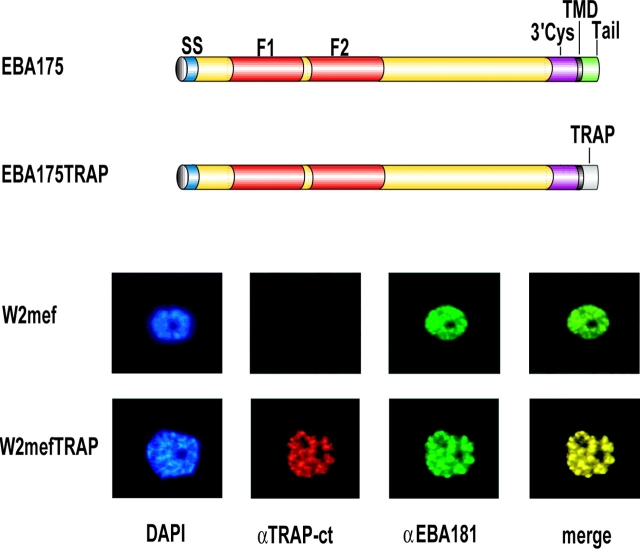
The invasion of host cells by the malaria parasite Plasmodium falciparum requires specific protein-protein interactions between parasite and host receptors and an intracellular translocation machinery to power the process. The transmembrane erythrocyte binding protein-175 (EBA-175) and thrombospondin-related anonymous protein (TRAP) play central roles in this process. EBA-175 binds to glycophorin A on human erythrocytes during the invasion process, linking the parasite to the surface of the host cell. In this report, we show that the cytoplasmic domain of EBA-175 encodes crucial information for its role in merozoite invasion, and that trafficking of this protein is independent of this domain. Further, we show that the cytoplasmic domain of TRAP, a protein that is not expressed in merozoites but is essential for invasion of liver cells by the sporozoite stage, can substitute for the cytoplasmic domain of EBA-175. These results show that the parasite uses the same components of its cellular machinery for invasion regardless of the host cell type and invasive form.


Figures

Figure 1.
Figure 1.
Sequence alignment of the cytoplasmic domain of microneme proteins and the EBA-175 mutations used in this paper. Also shown is the EBA-175 allelic replacement and Southern blot analysis. (A) Comparison of the microneme proteins EBA-175, EBA-181 (JESEBL), EBA-140 (BAEBL), EBL1, AMA1, TRAP, EBA165 (PAEBL), and MAEBL. The COOH-terminal amino acids of the highly conserved transmembrane domain are in black italics, tyrosine motifs and tyrosine residues are indicated by red letters, acidic amino acids are highlighted in blue. (B) Sequences of the constructs used in this paper to modify the cytoplasmic domain of EBA-175. COOH-terminal amino acids of the transmembrane domain are in black italics, and amino acid substitutions are in red. In gray is the sequence of TRAP fused to EBA-175 backbone. (C) Schematic representation of the 3′ replacement of the EBA-175 gene by single crossover recombination of pHH1 constructs in the EBA-175 locus. The positive selection cassette (hDHFR) of the pHH1 vector is represented by the black box. An ∼1.1-kb fragment of the COOH terminus with the introduced mutation (indicated by asterisks) was cloned in the pHH1 vector including the 3′ cysteine rich region (purple), the transmembrane domain (black), and the mutated cytoplasmic domain (green). This fragment is flanked by the 3′ UTR of the P. berghei dihydrofolate reductase gene (gray) in the pHH1 vector. Crosses refer to the regions where recombination events were expected. The intron/exon structure of the endogenous EBA-175 gene is shown. The red boxes indicate the adhesive F1/F2 ectodomains of the endogenous EBA-175. The MfeI (M) restriction sites are marked and the position of the EBA-175 probe used for Southern analysis is indicated. (D) Southern blot analysis of genomic DNA (MfeI restricted) of W2mef and transgenic EBA-175 mutant parasites reveals that the plasmid has integrated into the EBA-175 gene. Variable numbers of plasmid copies have integrated into each transgenic parasite. The position of the probe used in the Southern hybridization leads to large fragments of 8.1 and 11 kb that differ in intensity depending on the number of plasmids integrated. The 2.4-kb band is indicative of the integration of the plasmid via single recombination into the 3′ end of the EBA-175 gene. Importantly, the endogenous EBA-175 hybridizing band is 10 kb, and is different in the parasite lines where the 3′ end of EBA-175 has been replaced. Sizes of the hybridizing bands are shown in kb.
Figure 2.
Figure 2.
Immunoblot analysis of transgenic parasites expressing different EBA-175 mutant proteins. Proteins from synchronized parasite cultures were separated by SDS-PAGE on a 6% gel under reducing conditions. Proteins were detected with anti-EBA175, anti-EBA175-CT, anti-TRAP-CT, and anti-HSP70. Approximately equal signal was obtained with the anti-HSP70 antibodies, suggesting that each lane was loaded equally. In 3D7-derived parasite pellets, an additional cross-reactive band of ∼100 kD is detected by anti-EBA-175 antibodies. (B) Bar graph of the relative expression ratio of EBA-175 in late schizont stages of selected parasites. This ratio was calculated by dividing the average of three EBA175 assays with the average of the actin and histone 2B signals. The SD represents the sum of the EBA-175 and housekeeping SDs. The averages are calculated and the relative expression ratio is obtained by dividing the EBA-175 value with a mean of each control.
Figure 3.
Figure 3.
Immunolocalization of EBA-175 mutant proteins in transgenic parasites. The names on the left refer to the W2mef parasite lines expressing mutant EBA-175 proteins. The structure of EBA-175 and mutant proteins are schematically shown. Free merozoites were incubated with anti-EBA175 and anti-EBA181, followed by FITC-labeled anti–mouse and rhodamine-labeled anti–rabbit antibodies. To precisely visualize the localization of mutant EBA-175 with respect to the microneme protein EBA-181, the two fluorescence photomicrographs were merged. The function of the mutant EBA-175 was measured in each parasite line as shown in Fig. 4 A. + refers to absence of a switch in invasion demonstrating EBA-175 function is retained, whereas − signifies a switch in invasion phenotype and loss of function for EBA-175.
Figure 4.
Figure 4.
The deletion of the cytoplasmic domain of EBA-175 leads to a switch in the invasion phenotype. The TRAP cytoplasmic tail can reconstitute the function of the same region in EBA-175 for merozoite invasion. The ability to invade neuraminidase-treated erythrocytes is indicative of a switch in invasion to sialic acid independence and loss of EBA-175 function and this loss of function was confirmed directly by measurement of the ability to invade chymotrypsin-treated erythrocytes. RBCs were treated with neuraminidase (A) or chymotrypsin (B) as described in Materials and methods before testing in merozoite invasion assays. Figures shown are the percentage of invasion compared with untreated erythrocytes. Error bars correspond to standard deviation. The data obtained with neuraminidase-treated erythrocytes were from four independent experiments for W2mef, W2mef3′R and W2mefΔtail but only two independent experiments for W2mefTRAP. All experiments were performed in triplicate. The data obtained in panel B with chymotrypsin-treated erythrocytes represent one experiment done in triplicate. A second independent experiment has been performed and the results obtained are essentially the same as described here.
Figure 5.
Figure 5.
Expression and localization of EBA-175TRAP in transgenic parasites. Immunofluorescence assays using anti-TRAP-CT, anti-EBA-175 and anti-EBA-181 antibodies with parasite lines W2mef and W2mefTRAP are shown. Structural features of EBA-175 compared with the chimeric protein EBA-175TRAP. Nuclei are stained with DAPI in the first panel of each row. EBA-175 colocalizes with the microneme protein EBA-181 in W2mef parasites. EBA-175TRAP is not expressed in W2mef parasites but is expressed in W2mefTRAP transgenic parasites. It colocalizes with the micronemal protein EBA-181. The EBA-175 and EBA-181 patterns are merged to show colocalization in the last panel of each row.
Figure 6.
Figure 6.
A proposed model of trafficking and function for EBA-175. (A) Schematic representation of a merozoite. Secretory proteins enter the lumen of the endoplasmic reticulum (ER) with their NH2-terminal signal peptide. The molecular mechanism of pre- and post-Golgi trafficking (TGN) and the differential sorting of microneme (MIC) and rhoptry (R) proteins to their final destination is unclear. (B) In a post-Golgi sorting event, EBA-175 is trafficked to the micronemes by an escorter protein. The protein–protein interaction may take place via the conserved 3′ cysteine-rich region. The information of neither microneme localization nor interaction with the escorter protein is encrypted in the cytoplasmic domain of EBA-175. Deletion of the cytoplasmic domain does not affect sorting to the micronemes. Sorting was disrupted by deletion of the transmembrane domain and the 3′ cysteine rich region. (C) The intracellular function of EBA-175 is dependent on the interaction of an adaptor protein with the cytoplasmic domain. The deletion of this region abolishes the binding of the adaptor protein and leads to disruption of its function. The cytoplasmic domain can be substituted by the cytoplasmic domain of the sporozoite protein TRAP, suggesting that EBA-175 and TRAP have functionally equivalent roles in the active invasion process.

Similar articles

Cited by

References

    1. Adams, J.H., B.K.L. Sim, S.A. Dolan, X. Fang, D.C. Kaslow, and L.H. Miller. 1992. A family of erythrocyte binding proteins of malaria parasites. Proc. Natl. Acad. Sci. USA. 89:7085–7089. - PMC - PubMed
    1. Adams, J.H., P.L. Blair, O. Kaneko, and D.S. Peterson. 2001. An expanding ebl family of Plasmodium falciparum. Trends Parasitol. 17:297–299. - PubMed
    1. Aikawa, M., L.H. Miller, J. Johnson, and J. Rabbege. 1978. Erythrocyte entry by malarial parasites. A moving junction between erythrocyte and parasite. J. Cell Biol. 77:72–82. - PMC - PubMed
    1. Baldi, D.L., K.T. Andrews, R.S. Waller, D. Roos, B.S. Crabb, and A.F. Cowman. 2000. RAP1 controls rhoptry targeting of RAP2 in the malaria parasite Plasmodium falciparum. EMBO J. 19:1–9. - PMC - PubMed
    1. Barnwell, J.W., and M.R. Galinski. 1998. Invasion of vertebrate cells: erythrocytes. Malaria: Parasite Biology, Pathogenesis and Protection. I.W. Sherman, editor. ASM Press, Washington, DC. 93–120.

Publication types

MeSH terms