

Competitive binding of fatty acids and the fluorescent probe 1-8-anilinonaphthalene sulfonate to bovine beta-lactoglobulin
- PMID: 12876309
- PMCID: PMC2323946
- DOI: 10.1110/ps.0304403
Competitive binding of fatty acids and the fluorescent probe 1-8-anilinonaphthalene sulfonate to bovine beta-lactoglobulin
Abstract
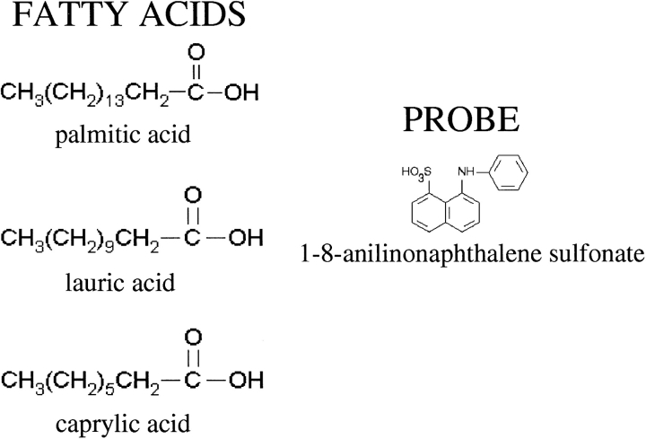
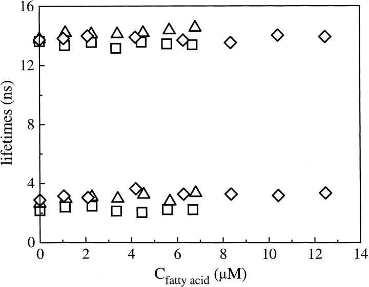
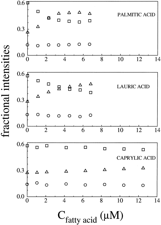
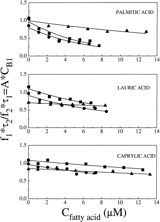
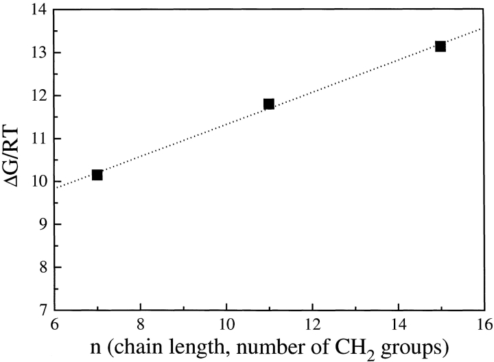
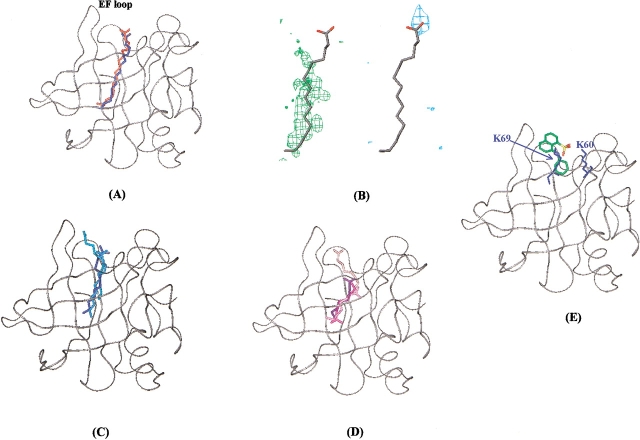
The use of spectroscopy in the study of fatty acids binding to bovine beta-lactoglobulin (BLG) appears to be a difficult task, as these acid compounds, assumed as the protein natural ligands, do not exhibit favorable optical response such as, for example, absorption or fluorescence. Therefore, the BLG fatty-acid equilibrium has been tackled by exploiting the competition between fatty acids and ANS, a widely used fluorescent hydrophobic probe, whose binding sites on the protein have been characterized recently. Two lifetime decays of the ANS-BLG complex have been found; the longer one has been attributed to the internal binding site and the shorter one to the external site. At increasing fatty acids concentration, the fractional weight associated with ANS bound to the internal site drops, in agreement with a model describing the competition of the dye with fatty acids, whereas the external site occupancy appears to be unaffected by the fatty acids binding to BLG. This model is supported by docking studies. An estimate of the acid-binding affinities for BLG has been obtained by implementing the fitting of the bound ANS intensities with a competitive binding model. A relevant dependence has been found upon the solution pH, in the range from 6 to 8, which correlates with the calyx accessibility modulated by the conformation of the EF loop. Fatty acids with longer aliphatic chains (palmitate and laurate) are found to display larger affinities for the protein and the interaction free energy nicely correlates with the number of contacts inside the protein calyx, in agreement with docking simulations.
Figures
References
-
- Anel, A., Calvo, M., Naval, J., Iturralde, M., Alava, M.A., and Piñeiro, A. 1989. Interaction of rat α-fetoprotein and albumin with polyunsaturated and other fatty acids: Determination of apparent association. FEBS Lett. 250 22–24. - PubMed
-
- Belitz, H.D. and Gosch, W. 1999. Food chemistry, p. 387. ed. Springer-Verlag, Heidelberg, Germany.
-
- Brownlow, S., Cabral, J.H.M., Cooper, R., Flower, D.R., Yewdall, S.J., Polikarpov, I., North, A.C.T., and Sawyer, L. 1997. Bovine β-lactoglobulin at 1.8 Å resolution—Still an enigmatic lipocalin. Structure 5 481–495. - PubMed
-
- Cho, Y., Batt, C.A., and Sawyer, L. 1994. Probing the retinol-binding site of bovine β-lactoglobulin. J. Biol. Chem. 269 11102–11107. - PubMed
-
- Collini, M., Chirico, G., Baldini, G., and Bianchi, M.E. 1995. Conformation of short DNA fragments by modulated fluorescence polarization anisotropy. Biopolymers 36 211–225. - PubMed
Publication types
MeSH terms
Substances
LinkOut - more resources
Full Text Sources
Molecular Biology Databases
