. 2003 Aug;47(8):2624-35.
doi: 10.1128/AAC.47.8.2624-2635.2003.

Entecavir therapy combined with DNA vaccination for persistent duck hepatitis B virus infection

Affiliations

Entecavir therapy combined with DNA vaccination for persistent duck hepatitis B virus infection

Wendy K Foster et al. Antimicrob Agents Chemother. 2003 Aug.

Abstract

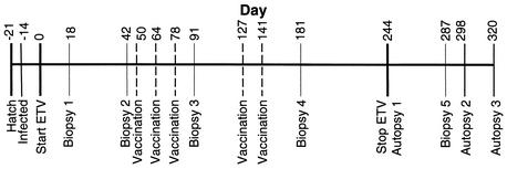
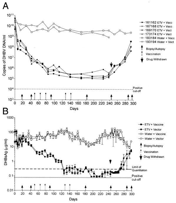
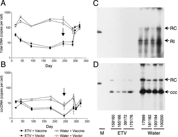
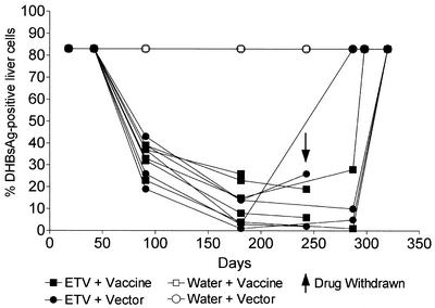
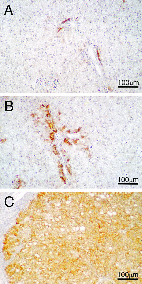
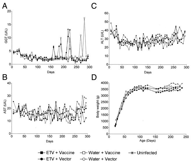
This study was designed to test the efficacy of antiviral treatment with entecavir (ETV) in combination with DNA vaccines expressing duck hepatitis B virus (DHBV) antigens as a therapy for persistent DHBV infection in ducks. Ducks were inoculated with 10(9) DHBV genomes at 7 days of age, leading to widespread infection of the liver and viremia within 7 days, and were then treated orally with either ETV (0.1 mg/kg of body weight/day) or distilled water from 21 days posthatch for 244 days. Treatment with ETV caused a 4-log drop in serum DHBV DNA levels within 80 days and a slower 2- to 3-log drop in serum DHBV surface antigen (DHBsAg) levels within 120 days. Following withdrawal of ETV, levels of serum DHBV DNA and DHBsAg rebounded to match those in the water-treated animals within 40 days. Sequential liver biopsy samples collected throughout the study showed that ETV treatment reduced DHBV DNA replicative intermediates 70-fold in the liver, while the level of the stable, template form, covalently closed circular DNA decreased only 4-fold. ETV treatment reduced both the intensity of antigen staining and the percentage of antigen-positive hepatocytes in the liver, but the intensity of antigen staining in bile duct cells appeared not to be effected. Intramuscular administration of five doses of a DNA vaccine expressing the DHBV presurface, surface, precore, and core antigens, both alone and concurrently with ETV treatment, on days 50, 64, 78, 127, and 141 did not result in any significant effect on viral markers.


Figures

FIG. 1.
FIG. 1.
Time line of the experiments performed in this study. Twenty-five ducks were divided into five treatment groups (1 to 5) containing five ducks each. The group 1 to 4 ducks were infected with DHBV at 7 days posthatch (day −14), while the group 5 ducks were used as uninfected controls. The group 1 and 2 ducks were treated daily with ETV for 244 days, while the group 3, 4, and 5 ducks were treated at the same time with distilled water. The group 1 and 3 ducks received DHBV vaccine, while the group 2 and 4 ducks received DNA vector on days 50, 64, 78, 127, and 141. All ducks underwent surgical liver biopsy on days 18, 42, 91, and 181. Two ducks from groups 1 to 4 (Table 1) were autopsied on day 243 (the day before drug was withdrawn), while the remaining ducks in groups 1 to 4 were biopsied on day 287 and then autopsied on either day 298 or day 320. The group 5 ducks were autopsied on day 320.
FIG. 2.
FIG. 2.
Detection of DHBV DNA (A) and DHBsAg (B) in the serum of ducks in groups 1 (ETV plus DHBV vaccine), 2 (ETV plus vector), 3 (water plus DHBV vaccine), and 4 (water plus vector). DHBV DNA was detected by real-time PCR using a Roche LightCycler in ducks 161162 and 167168 from group 1, 169170 and 173174 from group 2, 183184 from group 3, and 193194 from group 4. DHBsAg was detected by quantitative ELISA as described in the text, and the results for each group are shown as means ± standard deviations. Six ducks (one or two from each of groups 1 to 4 as described in the text) that had highly lipemic serum were excluded from analysis. The limit of quantitation (0.3 μg/ml) indicates the lower limit of the linear range of the standard curves, below which samples contained levels of DHBsAg that were still detectable but too low for accurate quantitation. The cutoff for DHBsAg-positive samples was set at 2 standard deviations above the average background obtained using uninfected duck serum.
FIG. 3.
FIG. 3.
Liver tissue was extracted for total and cccDNA and then subjected to Southern blot hybridization using a 32P-labeled genome-length DHBV DNA probe. (A and B) Graphs show the average total DHBV DNA (A) and DHBV cccDNA (B) levels per liver cell for all ducks in each group ± standard deviation. Arrows indicate when ETV treatment was withdrawn. (C and D) Liver tissue was collected from two ducks in each of groups 1 to 4 on day 243 after extraction for total DHBV DNA (C) and cccDNA (D) and Southern blot hybridization. Individual duck numbers are shown. Relaxed circular (RC) and cccDNA (ccc) were the predominant forms of DHBV DNA detected in the liver of the ETV-treated ducks on day 243. RI, replicative intermediates; M, 200 pg of plasmid DNA containing the 3-kb DHBV genome.
FIG. 4.
FIG. 4.
ETV treatment reduced the percentage of liver cells that contained detectable levels of DHBsAg. DHBsAg was detected in ethanol-acetic acid-fixed liver tissue from all ducks in groups 1 to 4 by using anti-DHBV pre-S monoclonal antibodies (1H.1 [28]) as described in the text. The percentage of liver cells expressing detectable levels of DHBsAg was determined by cell counts using a microscope grid as described in the text. Results from individual ducks are shown.
FIG. 5.
FIG. 5.
Detection of DHBsAg-positive cells in liver by indirect immunoperoxidase staining of ethanol-acetic acid-fixed liver tissue counterstained with hematoxylin from an ETV-treated duck from group 1 (167168) on day 287 (A), an ETV-treated duck from group 2 (173174) on day 287 in which DHBsAg detection in bile duct cells was largely unaffected by ETV treatment (B), and a water-treated DHBV-infected duck from group 4 (193194) on day 243 (C). Final magnification, ×114.
FIG. 6.
FIG. 6.
(A to C) Average levels of the liver function enzymes GGT (A), AST (B), and ALT (C) in serum collected at weekly intervals for all ducks in groups 1 to 5. (D) Average body weights for all ducks in each group are shown at weekly intervals.

References

    1. Bock, C. T., P. Schranz, C. H. Schroder, and H. Zentgraf. 1994. Hepatitis B virus genome is organized into nucleosomes in the nucleus of the infected cell. Virus Genes 8:215-229. - PubMed
    1. Chang, K. M., and F. V. Chisari. 1999. Immunopathogenesis of hepatitis B virus infection. Clin. Liver Dis. 3:221-239. - PubMed
    1. Chisari, F. V., and C. Ferrari. 1997. Viral hepatitis, p. 745-778. In N. Nathanson (ed.), Viral pathogenesis. Lippincott-Raven Publishers, Philadelphia, Pa.
    1. Colonno, R. J., E. V. Genovesi, I. Medina, L. Lamb, S. Durham, M. L. Huang, L. Corey, M. Littlejohn, S. Locarnini, B. C. Tennant, B. Rose, and J. M. Clark. 2001. Long-term entecavir treatment results in sustained antiviral efficacy and prolonged life span in the woodchuck model of chronic hepatitis infection. J. Infect. Dis. 184:1236-1245. - PubMed
    1. Fairbrother, A., M. A. Craig, K. Walker, and D. O'Loughlin. 1990. Changes in mallard (Anas platyrhynchos) serum chemistry due to age, sex, and reproductive condition. J. Wildl. Dis. 26:67-77. - PubMed

Publication types

MeSH terms

LinkOut - more resources