. 2003 Jul 23;23(16):6399-403.
doi: 10.1523/JNEUROSCI.23-16-06399.2003.

Identification of a Pax6-dependent epidermal growth factor family signaling source at the lateral edge of the embryonic cerebral cortex

Affiliations

Identification of a Pax6-dependent epidermal growth factor family signaling source at the lateral edge of the embryonic cerebral cortex

Stavroula Assimacopoulos et al. J Neurosci. .

Abstract

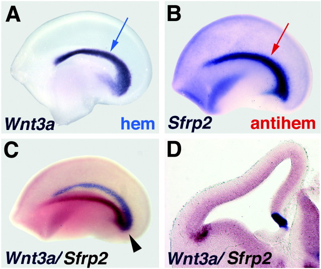
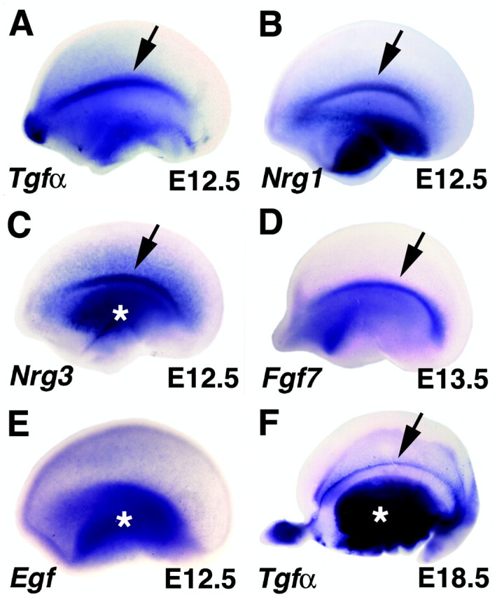
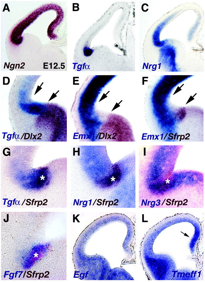
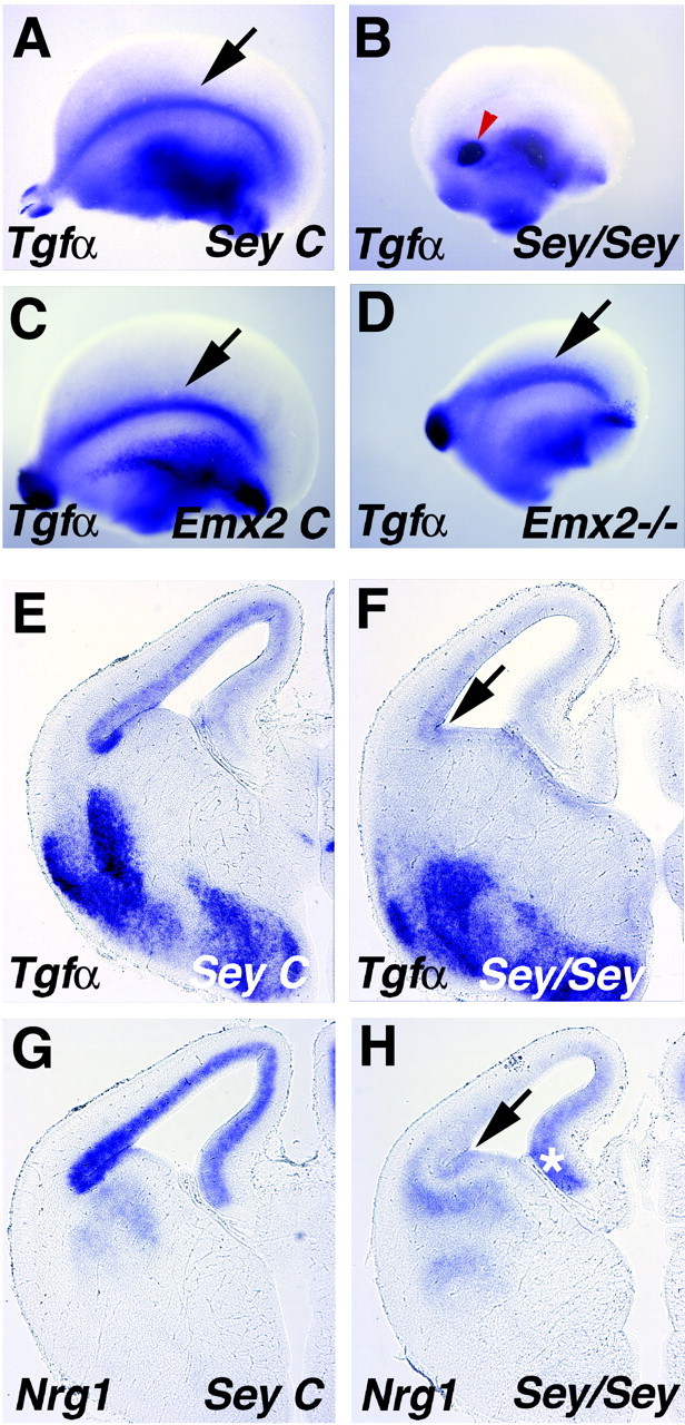
In an emerging model, area patterning of the mammalian cerebral cortex is regulated in part by embryonic signaling centers. Two have been identified: an anterior telencephalic source of fibroblast growth factors and the cortical hem, a medial structure expressing winglessint (WNT) and bone morphogenetic proteins. We describe a third signaling source, positioned as a mirror image of the cortical hem, along the lateral margin of the cortical primordium. The cortical antihem is identified by gene expression for three epidermal growth factor (EGF) family members, Tgf(alpha), Neuregulin 1, and Neuregulin 3, as well as two other signaling molecules, Fgf7 and the secreted WNT antagonist Sfrp2. We find that the antihem is lost in mice homozygous for the Small eye (Pax6) mutation and suggest the loss of EGF signaling at least partially explains defects in cortical patterning and cell migration in Small eye mice.


Figures

Figure 1.
Figure 1.
Location of the cortical hem and antihem. A-C, E12.5 cerebral hemispheres, processed with one- or two-color in situ hybridization and viewed from the medial (A) or lateral (B, C) faces. Anterior to the left. D, Coronal section through E12.5 cerebral hemisphere processed with two-color in situ hybridization. A, B, The “swoosh” of the medial cortical hem (A, blue arrow) is mirrored by the lateral antihem (B, red arrow). Together, they form a pincer arrangement around the cortical primordium (C). The cortical hem is marked by strong Wnt3a expression (purple in A, C,D), and the antihem is identified by expression of Sfrp2 (purple in B; reddish-brown in C, D). Arrowhead (C) indicates meeting of the hem and antihem in the caudal cerebral hemisphere.
Figure 2.
Figure 2.
EGF family members are expressed in the cortical antihem. A-E, Embryonic cerebral hemispheres viewed from the lateral face; anterior to the left (A-C, E, E12.5; D, E13.5). F, E18.5 hemisphere viewed from the inside looking laterally. A-C, Peaks of expression of Tgfα, Nrg1, and Nrg3 mark the curving longitudinal lateral band of the antihem (arrows in A-C). Tgfα expression is maintained for several days in this position (arrow, F). D, Fgf7 is also expressed in the antihem at E13.5. E, The founding member of the EGF family, Egf itself, is expressed in the ventral telencephalon and in the cortical primordium without a peak of expression at the antihem. Asterisks mark expression in ventral telencephalon (C, E, F).
Figure 3.
Figure 3.
Location of the antihem relative to the transition between dorsal and ventral telencephalon. A-L, Coronal sections through E12.5 and E13.5 (J) cerebral hemispheres processed for one- or two-color in situ hybridization. A, Ngn2 expression marks the boundaries of cortical neuroepithelium. B, The Tgfα-expressing domain lies within the cortical primordium (compare A, B). C, Nrg1 expression peaks in the Tgfα-rich domain but extends as a decreasing gradient into dorsolateral cortical neuroepithelium, with an additional zone of expression in the medial cortical primordium. D-F, At E12.5, a wedge-shaped territory lacks expression of Emx1 or Dlx2 (between arrows). This territory is filled by Tgfα expression (D), with Sfrp2 and Fgf7 expression nested in the Tgfα domain (F, G, J). Tgfα, Fgf7, and Sfrp2 expression remain within cortical neuroepithelium at this site (D, F, J), but Nrg1 and Nrg3 extend into the VZ of the basal telencephalon (H, I). Asterisks (G-J) mark the Sfrp2-expressing zone, the most lateral margin of the cortical neuroepithelium. K, L, At E12.5, Egf (K) and Tmeff1 (L), like other EGF family members, do not show gene expression peaks in the cortical antihem. Arrow (L) notes increased Tmeff1 expression in medial embryonic cortex, as seen in Nrg1-labeled sections (C).
Figure 4.
Figure 4.
The antihem is lost in the Small eye mouse. A-D, Cerebral hemispheres viewed from the lateral face; anterior to the left. E-H, Coronal sections through E14.5 cerebral hemispheres. A, B, E, F, The antihem marked by Tgfα expression is missing in an E14.5 mouse homozygous for the Small eye mutation (B, arrow in F) but is present in a littermate control (arrow in A, E). The spot of Tgfα labeling in Sey homozygote mice (red arrowhead in B) is likely the olfactory bulb remnant (Jimenez et al., 2000). C, D,A Tgfα-expressing antihem is present in an E13.5 Emx2 homozygote mutant mouse (D, arrow) and littermate control (C, arrow) but appears dorsally displaced in the mutant (D). G, H, Nrg1 expression is missing from the antihem region (arrow in H) and dorsolateral cortical neuroepithelium in an Sey/Sey mouse at E14.5, but expression in the hippocampal primordium remains (asterisk).

References

    1. Anderson SA, Marin O, Horn C, Jennings K, Rubenstein JL ( 2001) Distinct cortical migrations from the medial and lateral ganglionic eminences. Development 128: 353-363. - PubMed
    1. Bayer SA, Altman J, Russo RJ, Dai XF, Simmons JA ( 1991) Cell migration in the rat embryonic neocortex. J Comp Neurol 307: 499-516. - PubMed
    1. Bishop KM, Goudreau G, O'Leary DD ( 2000) Regulation of area identity in the mammalian neocortex by Emx2 and Pax6. Science 288: 344-349. - PubMed
    1. Caric D, Raphael H, Viti J, Feathers A, Wancio D, Lillien L ( 2001) EGFRs mediate chemotactic migration in the developing telencephalon. Development 128: 4203-4216. - PubMed
    1. Chapouton P, Gartner A, Gotz M ( 1999) The role of Pax6 in restricting cell migration between developing cortex and basal ganglia. Development 126: 5569-5579. - PubMed

Publication types

MeSH terms

Substances

LinkOut - more resources