. 2003 Jul 23;23(16):6413-22.
doi: 10.1523/JNEUROSCI.23-16-06413.2003.

Gap junctions mediate bystander cell death in developing retina

Affiliations

Gap junctions mediate bystander cell death in developing retina

Karen Cusato et al. J Neurosci. .

Abstract

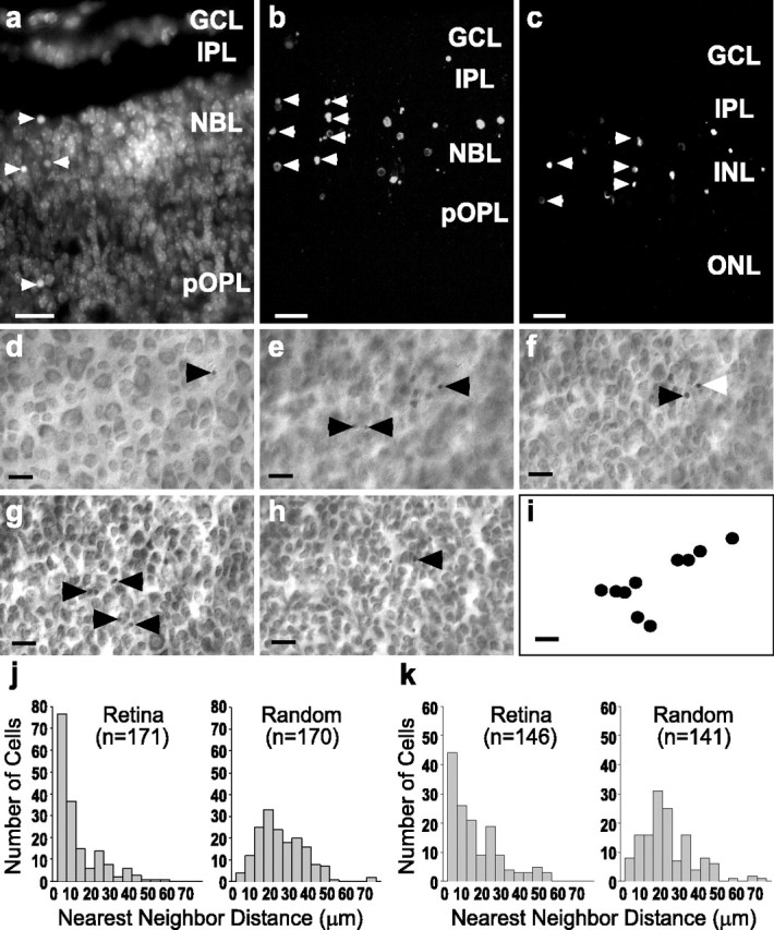
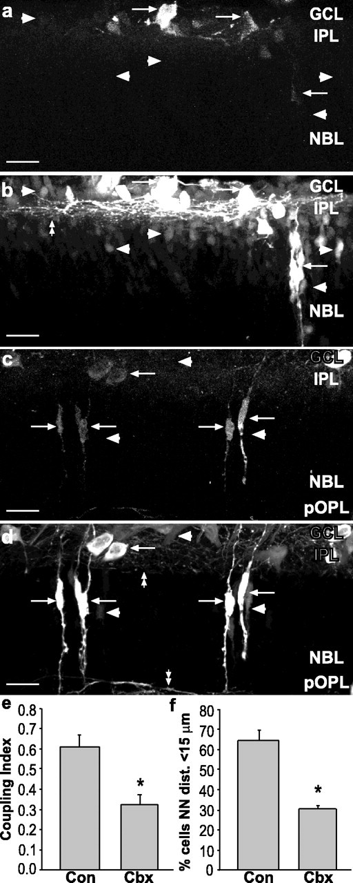
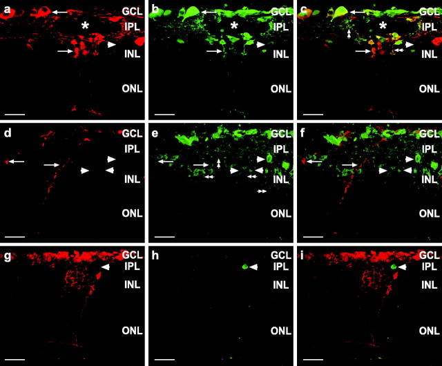
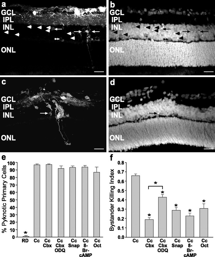
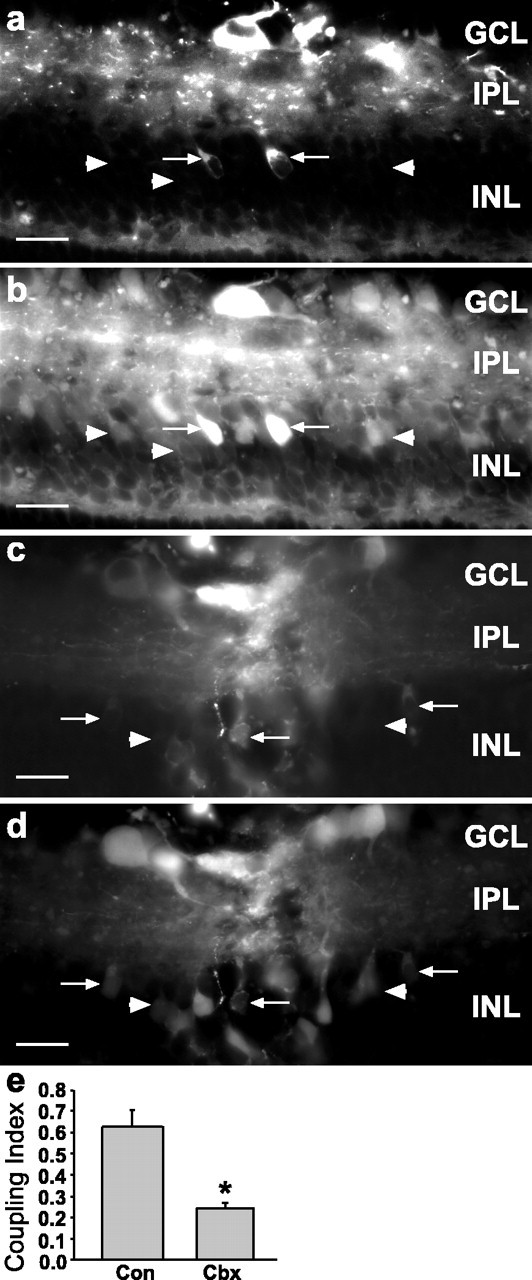
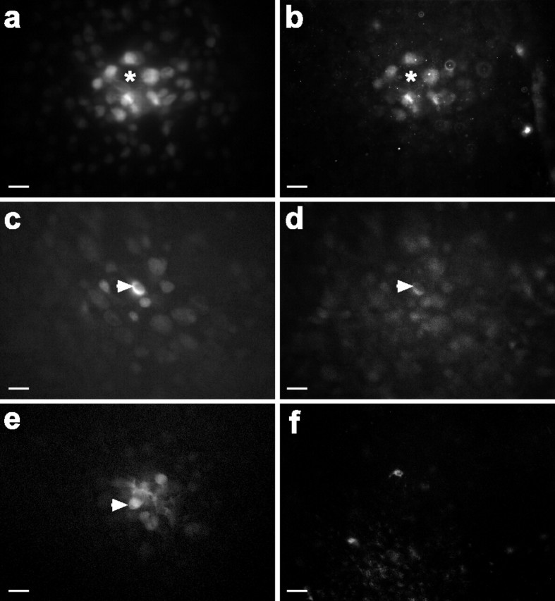
During development of the retina, programmed cell death helps to establish the final size and distribution of various cell classes in distinct layers of the tissue. Here we show that dying cells in the developing ganglion and inner nuclear layers are clustered spatially and that gap junction inhibitors decrease the clustering of dying cells. To confirm the role of gap junctions in cell death, we induced targeted cell death via intracellular cytochrome c (Cc) and examined the induced cells and their neighbors for apoptotic morphology or caspase-3 cleavage. These studies indicate that bystander killing extends to coupled cells. Quantitative studies of bystander killing were performed by scrape-loading retinas with Cc in the presence of rhodamine dextran (RD; to identify Cc-loaded cells) and by counting pyknotic cells in cryosections. Although only 1.5% of control scrape-loaded cells (RD alone) showed apoptotic morphology, 97% of Cc scrape-loaded cells were pyknotic. Moreover, bystander killing extended to neighboring cells, not labeled with RD, and was reduced significantly by the gap junction inhibitors octanol and carbenoxolone. We hypothesize that dying cells in the retina generate a gap junction-permeant apoptotic signal that mediates bystander killing. This novel finding of naturally occurring bystander cell death may have important implications in the histogenesis and pathology of the nervous system.


Figures

Figure 1.
Figure 1.
Spatial analysis of dying cells in the developing retina revealed a clustered distribution. a, Dying cells (arrowheads) evidenced by nuclear condensation (pyknosis) in DAPI-stained radial sections (20 μm) of P5 mouse retina. Z-series projections (15 μm) of TUNEL-labeled P5 (b) and P8 (c) mouse retina show that clusters of dying cells are frequent in developing retina. Scale bars in a-c, 10 μm. GCL, Ganglion cell layer; INL, inner nuclear cell layer; IPL, inner plexiform layer; NBL, neuroblastic layer; ONL, outer nuclear layer; OPL, outer plexiform layer; pOPL, presumptive OPL. d-i, Single field from a P5 mouse retina stained with cresyl violet, whole-mounted, and observed at different focal planes. Arrowheads point to pyknotic cells, and the white arrowhead indicates a pyknotic nucleus visible in multiple planes. d, Ganglion cell layer. e, Inner plexiform layer. f, g, Inner portion and outer portion (h) of inner nuclear layer. i, A two-dimensional surface reconstruction of the positions of the dying cells throughout the GCL and INL denotes the tendency of the dying cells to be clustered. Scale bar, d-i, 20 μm. j, Nearest neighbor (NN) distributions of P5 mouse retina (left) and of random points in fields of equal size and density (right). k, NN distribution of P8 mouse retina (left) and NN distribution of random points in fields of equal size and density (right). At both ages more dying cells are located near to one another in the retinas than in the random fields. All retinal NN distributions were significantly different from the associated random field NN distributions (Kolmogorov-Smirnov test, p < 0.001).
Figure 2.
Figure 2.
Carbenoxolone treatment decreased dye coupling and clustering of dying cells in vivo. a, b, Z-series projection (20 μm) of retinal sections from P5 mouse injected with vehicle and scrape-loaded with RD and NB. a, RD labels cells in GCL and NBL (arrows). b, NB (Cy2-streptavidin) is localized in cells that were scrape-loaded with RD (arrows) and other cells not loaded with RD (arrowheads). Many processes in the IPL were labeled with NB (double arrowhead). Arrowheads in a indicate position of coupled cells in b. c, d, Carbenoxolone treatment decreases spread of NB. c, RD labeling (arrows) of scrape-loaded retina from treated animal (20 mg/kg, s.c.). d, NB labeling of same field shows few NB-labeled cells that do not colocalize with RD (arrowheads). Processes in IPL and pOPL are indicated by double arrowheads. Scale bars in a-d, 20 μm. e, Coupling Index for control (Con) and carbenoxolone (Cbx) indicates a significant decrease in dye coupling by Cbx treatment (asterisks indicate groups significantly different from controls; Student's t test, p < 0.01). f, Cbx reduces the percentage of dying cells with a NN distance shorter than 15 μm in a manner that closely parallels the decrease in the Coupling Index.
Figure 3.
Figure 3.
Targeted killing of primary cells induced bystander cell death in vitro. a-i, Confocal images of radial sections (20 μm) from P13-P15 rat retinas scrape-loaded with or without Cc in the presence of RD-dextran. a-c, Retina scrape-loaded with RD and biotin-Cc. The asterisks denote the shallow scrape-loading incision spanning the GCL and IPL in part of the z-series. Shown are RD (red, a), biotin-Cc (Cy2-streptavidin; green, b), and the merged image (c). RD and Cc are both too large to pass through gap junctions and thus remain confined to the scrape-loaded cells. Both somata (arrows) and processes (double arrowheads) are labeled. RD and Cc colocalize (c) with only occasional cells labeled for Cc alone (arrowhead); therefore, RD clearly identifies the cells loaded with Cc. d-f, Retina scrape-loaded with RD and Cc, immunolabeled for cleaved caspase-3. d, RD-labeled cells (red; arrows) were immunopositive (e) for cleaved caspase-3 (green; arrows). Cells not labeled with RD also were labeled for cleaved caspase-3 (arrowheads), indicating bystander cell death. Cleaved caspase-3 was detected in both somata and processes (double arrowheads). f, Merged image indicates that RD and cleaved caspase were colocalized (arrows) in some cells, but other dying cells did not contain RD (arrowheads). g-i, Control retina scrape-loaded with RD alone. g, RD-labeled cells (red) were not labeled for cleaved caspase-3 (h). However, one cell not labeled with RD was dying (h, i; arrowhead). This indicates that neither the scrape-loading nor the RD was a potent inducer of cell death. i, Merged image. Scale bars in a-i, 20 μm.
Figure 4.
Figure 4.
The propagation of cell death was decreased by the inhibition of gap junction coupling in vitro. a, b, Radial sections of P13 rat retina scrape-loaded with RD and Cc. RD labeling (a) identified the primary cells loaded with Cc, and DAPI staining (b) showed pyknotic nuclei in cells labeled with RD (arrows) and also in many bystander cells unlabeled with RD (arrowheads). Note in a and b that the RD-loaded horizontal cells (double arrowheads) were not pyknotic. c, d, Retina scrape-loaded with RD alone. c, RD-labeled cells were occasionally pyknotic (arrow) in DAPI staining (d) of the same field. Scale bars in a-d, 20 μm. e, Percentage of scrape-loaded cells killed for control (RD), Cc, and Cc with drug treatments. RD was significantly different from Cc (p < 0.01; one-way ANOVA, Duncan's multiple range test), but treatment with gap junction inhibitors did not decrease the death of cells loaded with Cc. f, The Bystander Killing Index for retinas treated with Cc demonstrates that gap junction inhibitors (carbenoxolone and octanol) as well as cAMP and NO generators (SNAP) decreased the bystander killing effect significantly when compared with Cc alone (asterisks indicate significant differences from Cc; p < 0.01; one-way ANOVA, Duncan's multiple range test). ODQ significantly attenuated the effects of carbenoxolone, indicating a role for cGMP as well. None of these agents decreased cell death in primary cells (e), indicating that the rescue effect was specific for the bystander cells. Cc, Cytochrome c; Cbx, carbenoxolone; ODQ, 1H-[1,2,4]oxadiazolo[4,3a]quinoxalin-1-one; SNAP, S-nitroso-dl-penicillamine; Oct, octanol.
Figure 5.
Figure 5.
Carbenoxolone decreased dye coupling in the scrape-loading model. Shown is control P13 rat retina scrape-loaded with RD (a) and Neurobiotin (b), which was visualized with Cy2-streptavidin. In a and b note that many Neurobiotin-labeled cells were not labeled with RD (arrowheads), indicating that they were coupled to the scrape-loaded cells (arrows). c, d, P13 retina treated with carbenoxolone (20 mg/kg, s.c., followed by 10 μm), scrape-loaded with RD and Neurobiotin. d, Neurobiotin labeling of same field as in c; only occasional cells were single labeled for Neurobiotin (arrowheads), indicating decreased coupling as compared with control (a, b). The same concentration of carbenoxolone that decreased bystander cell killing inhibited coupling to similar levels. Scale bars in a-d, 20 μm. e, The Coupling Index for retinas treated with carbenoxolone demonstrates that this gap junction inhibitor decreased the Coupling Index significantly as compared with control (asterisk indicates group significantly different from control; two-tailed t test, p < 0.01). Con, Control; Cbx, carbenoxolone.
Figure 6.
Figure 6.
Dying bystander cells were coupled via gap junctions to primary cells. a-d, Single-cell injections of AF488 (a gap junction-permeable dye) and Cc in the GCL showed that multiple coupled cells were immunolabeled for cleaved caspase-3. a, AF488 labeled cells coupled to the injected cell. Asterisks show location of injected cell that was no longer present 1 hr after the injection; presumably, it died and detached or was cleared by engulfment. b, Cleaved caspase-3 labeled many of the coupled cells in a. c, d, AF488 (c) and cleaved caspase-3 (d) labeling showed that the injected cell (arrowhead) and other coupled cells were immunopositive. e, f, Control injections of AF488 (e) into a single cell (arrowhead) did not induce caspase-3 cleavage (f) in the injected or coupled cells. Scale bars in a-f, 20 μm.

References

    1. Becker DL, Bonness V, Catsicas M, Mobbs P ( 2002) Changing patterns of ganglion cell coupling and connexin expression during chick retinal development. J Neurobiol 15: 280-293. - PubMed
    1. Brustugun OT, Fladmark KE, Doskeland SO, Orrenius S, Zhivotovsky B ( 1998) Apoptosis induced by microinjection of cytochrome c is caspase-dependent and is inhibited by Bcl-2. Cell Death Differ 5: 660-668. - PubMed
    1. Chang GQ, Hao Y, Wong F ( 1993) Apoptosis: final common pathway of photoreceptor death in rd, rds, and rhodopsin mutant mice. Neuron 11: 595-605. - PubMed
    1. Cook B, Lewis GP, Fisher SK, Adler R ( 1995) Apoptotic photoreceptor degeneration in experimental retinal detachment. Invest Ophthalmol Vis Sci 36: 990-996. - PubMed
    1. Cook B, Portera-Cailliau C, Adler R ( 1998) Developmental neuronal death is not a universal phenomenon among cell types in the chick embryo retina. J Comp Neurol 396: 12-19. - PubMed

Publication types