. 2003 Jul 23;23(16):6490-8.
doi: 10.1523/JNEUROSCI.23-16-06490.2003.

Long-term effects of permanent vestibular lesions on hippocampal spatial firing

Affiliations

Long-term effects of permanent vestibular lesions on hippocampal spatial firing

Noah A Russell et al. J Neurosci. .

Abstract

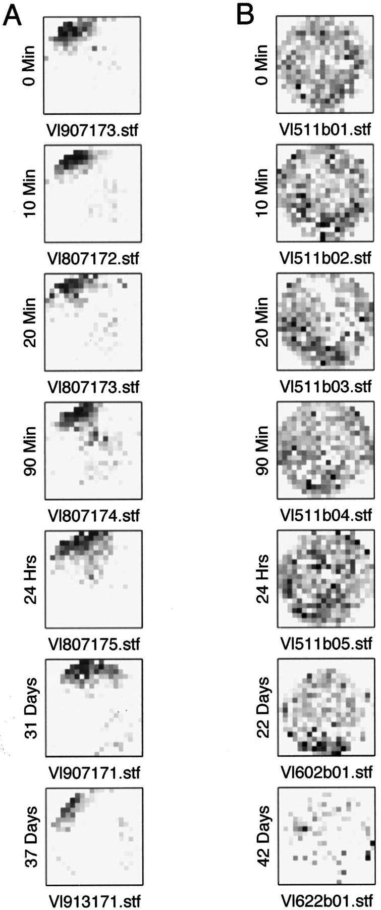
The hippocampus is thought to be important for spatial representation processes that depend on the integration of both self-movement and allocentric cues. The vestibular system is a particularly important source of self-movement information that may contribute to this spatial representation. To test the hypothesis that the vestibular system provides self-movement information to the hippocampus, rats were given either a bilateral labyrinthectomy (n = 6) or a sham surgery (n = 6), and at least 60 d after surgery hippocampal CA1 neurons were recorded extracellularly while the animals foraged freely in an open arena. Recorded cells were classified as complex spiking (n = 80) or noncomplex spiking (n = 33) neurons, and their spatial firing fields (place fields) were examined. The most striking effect of the lesion was that it appeared to completely abolish location-related firing. The results of this and previous studies provide converging evidence demonstrating that vestibular information is processed by the hippocampus. The disruption of the vestibular input to the hippocampus may interfere with the reconciliation of internal self-movement signals with the changes to the external sensory inputs that occur as a result of that movement. This would disrupt the ability of the animal to integrate allocentric and egocentric information into a coherent representation of space.


Figures

Figure 1.
Figure 1.
The recording protocols. All recordings consisted of blocks of 10 min sessions. CS cells were recorded for a single session (A), for five sessions with delays (AAA-A-A), or in a light-dark-light (ABA) protocol. Non-CS cells were only recorded for a single session. At least 5 min of habituation time was allowed to elapse before any recording was started.
Figure 2.
Figure 2.
Comparison of the behavior of control and lesioned animals. A, Mean velocity distributions over all control sessions (n = 171) and all lesion sessions (n = 193). Note that the lesioned animals spend a greater proportion of the time moving faster, reflecting their hyperactivity. The SE bars have been omitted from this figure because the values were so low that they interfered with the clarity of the symbols denoting the two groups. Despite their hyperactivity, the lesioned animals did not differ in their coverage of the environment during exploration (B) or in the heterogeneity of their exploration as assessed by the coefficient of variation of pixel dwell times (C).
Figure 3.
Figure 3.
Representative firing rate maps for CS cells in control (A) and lesion (B) animals. Note that the spatially dependent firing for the lesioned animals is less well defined. The fields shown were selected objectively by finding the field with properties that were closest to average.
Figure 4.
Figure 4.
Representative firing rate maps for non-CS cells in control (A) and lesion (B) animals. Although the spatial firing correlates are very weak for non-CS fields, they do exist. The fields shown were selected objectively by finding the field with properties that were the closest to average.
Figure 5.
Figure 5.
Firing rate map cross-correlations between sessions in the AAA-A-A protocol. Lesion maps had lower cross-correlations, indicating instability over a period of several minutes. A, Mean and SEM cross-correlations over all cells. B, Cross-correlations from one control and one lesion cell recorded for 6 weeks.
Figure 6.
Figure 6.
Firing rate maps for the cells recorded over a 6 week period shown in Figure 5B. A, Control CS cell. B, Lesion CS cell.
Figure 7.
Figure 7.
Changes in spatially dependent firing in the lesion group cells over time. A, Cross-correlations between the firing rate map recorded during the first 5 min block of a 30 min continuous recording session and each subsequent 5 min block. Lesion group maps have low cross-correlations immediately on the first comparison, indicating instability over a period of a few minutes. B, Mean cumulative place field size. Field size was calculated from firing data between t0 and a later time tn, where tn increased in 5 min increments from 5 to 30 min. Control group fields reach maximum size within 5 min with no further cumulative increase. In contrast, lesion group fields continue to increase in size with no obvious asymptote. Collectively these results suggest that there is no spatially dependent firing in the lesioned animals.
Figure 8.
Figure 8.
Firing rate maps recorded in the light-dark-light (ABA) protocol in control (A) and lesion (B) animals. The changes to the properties of the firing field that occurred in the dark were no greater than the changes in the light, over the same time period, for either control or lesioned animals. Hence the observed changes to the lesion animal's field in the dark cannot be attributed to the absence of vision.
Figure 9.
Figure 9.
Potential polysynaptic anatomical pathways from the VNC to the hippocampus. The thalamocortical route is shown on the left, and the septohippocampal and head direction system routes are shown on the right for clarity only. The pathways have been drawn onto a scale two-dimensional topographical projection of the rat brain. The different shaded regions indicate, from bottom to top, the myencephalon, metencephalon, mesencephalon, diencephalon, and telencephalon, respectively. VNC, Vestibular nucleus complex; Prh, perirhinal cortex; EC, entorhinal cortex; PPTN, pendunculopontine tegmental nucleus; Sum, supramammillary nucleus; MS, medial septum; DTN, dorsal tegmental nucleus; LMN, lateral mammillary nucleus; AND, anterodorsal thalamic nucleus; Post, postsubiculum.

Similar articles

Cited by

References

    1. Bannerman DM, Gilmour G, Norman G, Lemaire M, Iversen SD, Rawlins JN ( 2001) The time course of the hyperactivity that follows lesions or temporary inactivation of the fimbria-fornix. Behav Brain Res 120: 1-11. - PubMed
    1. Basile AS, Brichta AM, Harris BD, Morse D, Coling D, Skolnick P ( 1999) Dizocilpine attenuates streptomycin-induced vestibulotoxicity in rats. Neurosci Lett 265: 71-74. - PubMed
    1. Berger TW, Rinaldi PC, Weisz DJ, Thompson RF ( 1983) Single-unit analysis of different hippocampal cell types during classical conditioning of rabbit nictitating membrane response. J Neurophysiol 50: 1197-1219. - PubMed
    1. Bilkey DK, Muir GM ( 1999) A low cost, high precision subminiature microdrive for extracellular unit recording in behaving animals. J Neurosci Methods 92: 87-90. - PubMed
    1. Bilkey DK, Russell NA, Colombo M ( 2003) A lightweight microdrive for single unit recording in freely moving rats and pigeons. Methods 30: 152-158. - PubMed

Publication types

LinkOut - more resources