. 2003 Aug 5;100(16):9614-9.
doi: 10.1073/pnas.1531957100. Epub 2003 Jul 23.

Estrogen receptor-alpha mediates the brain antiinflammatory activity of estradiol

Affiliations

Estrogen receptor-alpha mediates the brain antiinflammatory activity of estradiol

Elisabetta Vegeto et al. Proc Natl Acad Sci U S A. .

Abstract

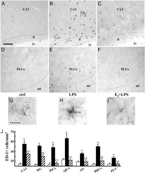
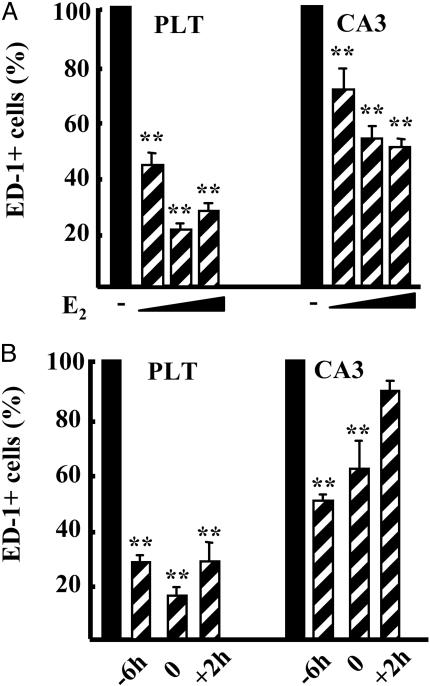
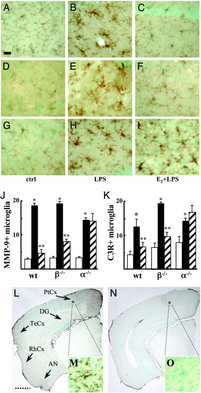
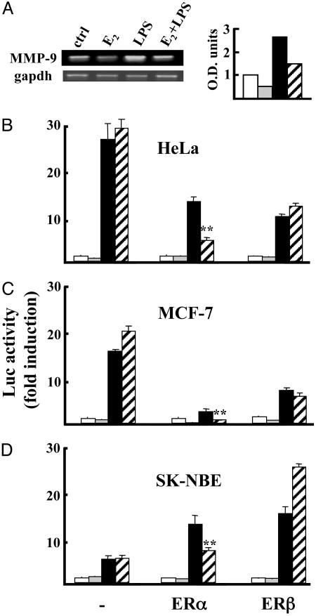
Beyond the key role in reproductive and cognitive functions, estrogens have been shown to protect against neurodegeneration associated with acute and chronic injuries of the adult brain. Current hypotheses reconcile this activity with a direct effect of 17beta-estradiol (E2) on neurons. Here we demonstrate that brain macrophages are also involved in E2 action on the brain. Systemic administration of hormone prevents, in a time- and dose-dependent manner, the activation of microglia and the recruitment of peripheral monocytes induced by intraventricular injection of lipopolysaccharide. This effect occurs by limiting the expression of neuroinflammatory mediators, such as the matrix metalloproteinase 9 and lysosomal enzymes and complement C3 receptor, as well as by preventing morphological changes occurring in microglia during the inflammatory response. By injecting lipopolysaccharide in estrogen receptor (ER)-null mouse brains, we demonstrate that hormone action is mediated by activation of ERalpha but not of ERbeta. The specific role of ERalpha is further confirmed by comparing the effects of ERs on the matrix metalloproteinase 9 promoter activity in transient transfection assays. Finally, we report that genetic ablation of ERalpha is associated with a spontaneous reactive phenotype of microglia in specific brain regions of adult ERalpha-null mice. Altogether, these results reveal a previously undescribed function for E2 in brain and provide a mechanism for its beneficial activity on neuroinflammatory pathologies. They also underline the key role of ERalpha in brain macrophage reactivity and hint toward the usefulness of ERalpha-specific drugs in hormone replacement therapy of inflammatory diseases.


Figures

Fig. 1.
Fig. 1.
Estrogen prevents activation of brain macrophages induced by LPS. Shown are representative immunohistochemical assays with ED-1 as a marker for microglia and monocytes in the CA3 field of the hippocampus (A–C) and in posterolateral cortical amygdaloid nuclei (DF); animals were injected with saline (A and D), LPS (B and E), or E2 before LPS (C and F). CA3, CA3 field of the hippocampus; fi, fimbria; 3v, third ventricle. PLCo, posterolateral cortical amygdaloid nuclei; ML, molecular layer. (Bar = 35 μm.) (G–I) Isolectin-B4 was used to visualize microglial morphology in the CA3 region of animals treated as specified. (Dashed bar = 8 μm.) (J) The number of ED-1-positive microglial cells was counted in the CA3 and DG of the hippocampus, in the parietal cortex (PtCx), cingulate cortex (cgCx), amygdaloid nuclei (AN), rhinal cortex (RhCx), and posterolateral thalamic nuclei (PLT). Saline, LPS, and E2 + LPS treatments are represented as open, filled, and dashed bars, respectively. Values are the mean ± SD of cells counted in adjacent sections from a single representative experiment, repeated at least three times. *, P < 0.01 vs. control; **, P < 0.01 vs. LPS.
Fig. 2.
Fig. 2.
Time- and dose-dependent inhibitory effect of estrogen on brain macrophage activation. (A) The number of ED-1-positive cells in the posterolateral thalamus (PLT) and CA3 was counted in rat brains injected with LPS 6 h after s.c. injection of vehicle (filled bars) or increasing doses of E2 (0.5, 5, and 50 μg/kg of E2; dashed bars). (B) Fifty micrograms/kilogram E2 injected 6 h before (-6h), at the same time as (0), or 2 h after (+2h) LPS. Bars are the average ± SD of a single representative experiment made on three animals; the experiment was repeated three times. Data are calculated as percentage of LPS alone. **, P < 0.01 vs. LPS.
Fig. 3.
Fig. 3.
ER-α mediates the inhibitory activity of E2 on microglia activation. MMP-9 expression in the CA1 region of wild-type (A–C), ERβ-null (D–F), and ERα-null (G–I) mice injected icv with saline (A, D, and G), LPS (B, E, and H), or E2 before LPS (C, F, and I). (J and K) Quantification of estrogen activity on the number of MMP-9-positive (J) and C3R-positive (K) cells per mm2 in the CA1 region of mice with different ER genetic background. Saline, LPS, and E2 + LPS treatments are represented as open, filled, and dashed bars, respectively. Bars are from a single representative experiment, repeated at least three times. Values represent the mean ± SD of cells counted in different adjacent sections. *, P < 0.01 vs. control; **, P < 0.01 vs. LPS. (L–O) Disruption of ERα results in brain microglia activation. Microglial cells were visualized by immunohistochemistry of C3R on brain coronal sections of ERα-null mice at 4 mo (L and M) or 2 mo (N and O) of age. (L) Strong C3R staining shows a reactive phenotype in the parietal cortex (PtCx), DG, temporal cortex (TeCx), rhinal cortex (RhCx), and amygdaloid nuclei (AN; see arrows). (M and O) Enlargements showing the reactive phenotype of microglia only in a 4-mo-old ERα-null mouse. Sections were counterstained with methyl green. (Filled bars = 35 μm, dashed bars = 1.2 mm, and open bars = 7 μm.)
Fig. 4.
Fig. 4.
E2-activated ER-α inhibits the induction of MMP-9 expression. (A) RT-PCR detection of MMP-9 RNA levels in the brain. (Right) Optical density of the bands as normalized with GAPDH. Groups of three animals were treated with saline (ctrl, open bar), E2 alone (light gray bar), LPS (filled bar), or E2 + LPS (dashed bar). Data are from single animals representative of each experimental group; the experiment was repeated three times. (B) MMP-9 promoter regulation by ERs. HeLa (B), MCF-7 (C), and SK-NBE (D) cell lines were transfected with the MMP-9-luc promoter alone (-) or with ERα or ERβ and treated with vehicle (open bars), 10-9 M E2 (light gray bars), 10-8 M phorbol 12-myristate-13-acetate (PMA; filled bars), or E2+PMA (dashed bars). Values represent the mean ± SD of triplicate samples of a single experiment repeated at least three times. *, P < 0.01 vs. LPS.

Similar articles

Cited by

References

    1. Sherwin, B. (2002) Trends Pharmacol. Sci. 23, 527-534. - PubMed
    1. Freeman, M. P., Smith, K. W., Freeman, S. A., McElroy, S. L., Kmetz, G. E., Wright, R. & Keck, P. E., Jr. (2002) J. Clin. Psychiatry 63, 284-287. - PubMed
    1. Huber, T. J., Rollnik, J., Wilhelms, J., von zur Muhlen, A., Emrich, H. M. & Schneider, U. (2001) Psychoneuroendocrinology 26, 27-35. - PubMed
    1. Henderson, W. (1997) Neurology 48, S27-S35. - PubMed
    1. Yaffe, K., Sawaya, G., Lieberburg, I. & Grady, D. (1998) J. Am. Med. Assoc. 9, 688-695. - PubMed

Publication types

MeSH terms