

An unusual retinal vascular morphology in connection with a novel AIPL1 mutation in Leber's congenital amaurosis
- PMID: 12881340
- PMCID: PMC1771788
- DOI: 10.1136/bjo.87.8.980
An unusual retinal vascular morphology in connection with a novel AIPL1 mutation in Leber's congenital amaurosis
Abstract
Aims: To report a case of an unusual retinal vascular morphology in connection with a novel AIPL1 mutation in a patient with Leber's congenital amaurosis (LCA).
Methods: A patient with LCA and no light perception from birth had both eyes enucleated at the age of 22 years because of excruciating pain. Mutation analysis was performed on known LCA genes. The eyes were processed for casts of the vascular tree, routine histopathology, and electron microscopy.
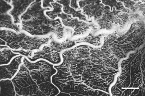
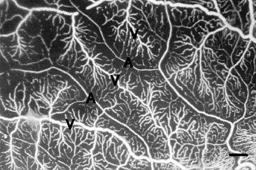
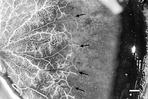
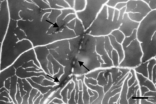
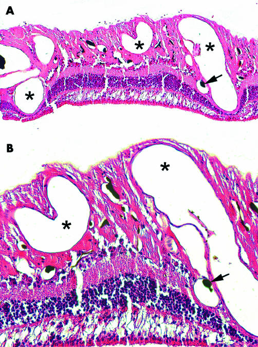
Results: A novel H82Y (244C-->T) mutation and a H90D (286G-->C) polymorphism were detected in exon 2 of the AIPL1 gene. Both the cast and the histopathological examination showed dilated retinal vessels, mainly venules, primarily localised in the posterior pole. In the mid-peripheral retina the density of capillaries on the arteriolar side of the microcirculatory units was significantly decreased. The vascular system was seen to gradually attenuate towards the retinal periphery, and to stop at a zone located approximately 4 mm from the ora serrata along the whole circumference. In this zone pigmented aggregates characteristic of retinitis pigmentosa were seen to ensheath the retinal vessels. The photoreceptors were almost totally absent and retinal gliosis was present. A decreased number of ganglion cells and an increased vacuolisation of the nerve fibre layer were observed. The retinal pigment cells and Bruch's membrane appeared normal in all regions.
Conclusion: An unusual retinal vascular morphology in an LCA patient is presented and possible pathogenic mechanisms of the findings are discussed.
Figures
Similar articles
-
Predominant rod photoreceptor degeneration in Leber congenital amaurosis.Mol Vis. 2005 Jul 22;11:542-53. Mol Vis. 2005. PMID: 16052170
-
Leber's congenital amaurosis with anterior keratoconus in Pakistani families is caused by the Trp278X mutation in the AIPL1 gene on 17p.Can J Ophthalmol. 2001 Aug;36(5):252-9. doi: 10.1016/s0008-4182(01)80018-1. Can J Ophthalmol. 2001. PMID: 11548141
-
Prenatal human ocular degeneration occurs in Leber's congenital amaurosis (LCA2).J Gene Med. 2002 Jul-Aug;4(4):390-6. doi: 10.1002/jgm.278. J Gene Med. 2002. PMID: 12124981
-
[Past, present, and future in Leber's hereditary optic neuropathy].Nippon Ganka Gakkai Zasshi. 2001 Dec;105(12):809-27. Nippon Ganka Gakkai Zasshi. 2001. PMID: 11802455 Review. Japanese.
-
[From gene to disease; Leber congenital amaurosis (LCA)].Ned Tijdschr Geneeskd. 2005 Oct 15;149(42):2334-7. Ned Tijdschr Geneeskd. 2005. PMID: 16261712 Review. Dutch.
Cited by
-
Clinical and genetic characteristics of Leber congenital amaurosis with novel mutations in known genes based on a Chinese eastern coast Han population.Graefes Arch Clin Exp Ophthalmol. 2016 Nov;254(11):2227-2238. doi: 10.1007/s00417-016-3428-5. Epub 2016 Jul 16. Graefes Arch Clin Exp Ophthalmol. 2016. PMID: 27422788
-
Metabolic Signaling in a Theoretical Model of the Human Retinal Microcirculation.Photonics. 2021 Oct;8(10):409. doi: 10.3390/photonics8100409. Epub 2021 Sep 23. Photonics. 2021. PMID: 36052288 Free PMC article.
-
Do photoreceptor cells cause the development of retinal vascular disease?Vision Res. 2017 Oct;139:65-71. doi: 10.1016/j.visres.2017.03.011. Epub 2017 May 8. Vision Res. 2017. PMID: 28438678 Free PMC article. Review.
-
The Leber congenital amaurosis protein AIPL1 functions as part of a chaperone heterocomplex.Invest Ophthalmol Vis Sci. 2008 Jul;49(7):2878-87. doi: 10.1167/iovs.07-1576. Epub 2008 Apr 11. Invest Ophthalmol Vis Sci. 2008. PMID: 18408180 Free PMC article.
-
Photoreceptor cells and RPE contribute to the development of diabetic retinopathy.Prog Retin Eye Res. 2021 Jul;83:100919. doi: 10.1016/j.preteyeres.2020.100919. Epub 2020 Nov 12. Prog Retin Eye Res. 2021. PMID: 33188897 Free PMC article. Review.
References
-
- Smith D, Oestreicher J, Musarella MA. Clinical spectrum of Leber’s congenital amaurosis in the second to fourth decades of life. Ophthalmology 1990;97:1156–61. - PubMed
-
- Waardenburg PJ, Schappert-Kimmijser J. On various recessive biotypes of Leber’s congenital amaurosis. Acta Ophthalmol (Copenh) 1963;41:317–20. - PubMed
-
- Perrault I, Rozet JM., Calvas P, et al. Retinal-specific guanylate cyclase gene mutations in Leber’s congenital amaurosis. Nat Genet 1996;14:461–4. - PubMed
-
- Gu S-M, Thompson DA, Srikumari CRS, et al. Mutations in RPE65 cause autosomal recessive childhood-onset severe retinal dystrophy. Nat Genet 1997;17:194–7. - PubMed
-
- Freund CL, Wang QL, Chen S, et al. De novo mutations in the CRX homeobox gene associated with Leber congenital amaurosis. Nat Genet 1998;18:311–2. - PubMed
Publication types
MeSH terms
Substances
LinkOut - more resources
Full Text Sources
Research Materials
Miscellaneous