

Unilateral high myopia: optical components, associated factors, and visual outcomes
- PMID: 12881349
- PMCID: PMC1771811
- DOI: 10.1136/bjo.87.8.1025
Unilateral high myopia: optical components, associated factors, and visual outcomes
Abstract
Aim: To elucidate the optical basis for unilateral high myopia and to identify the factors associated with its development.
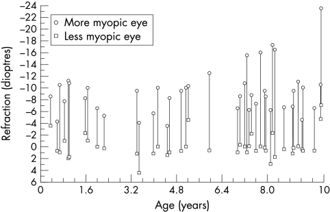
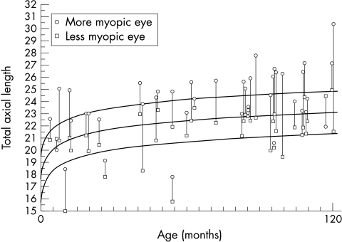
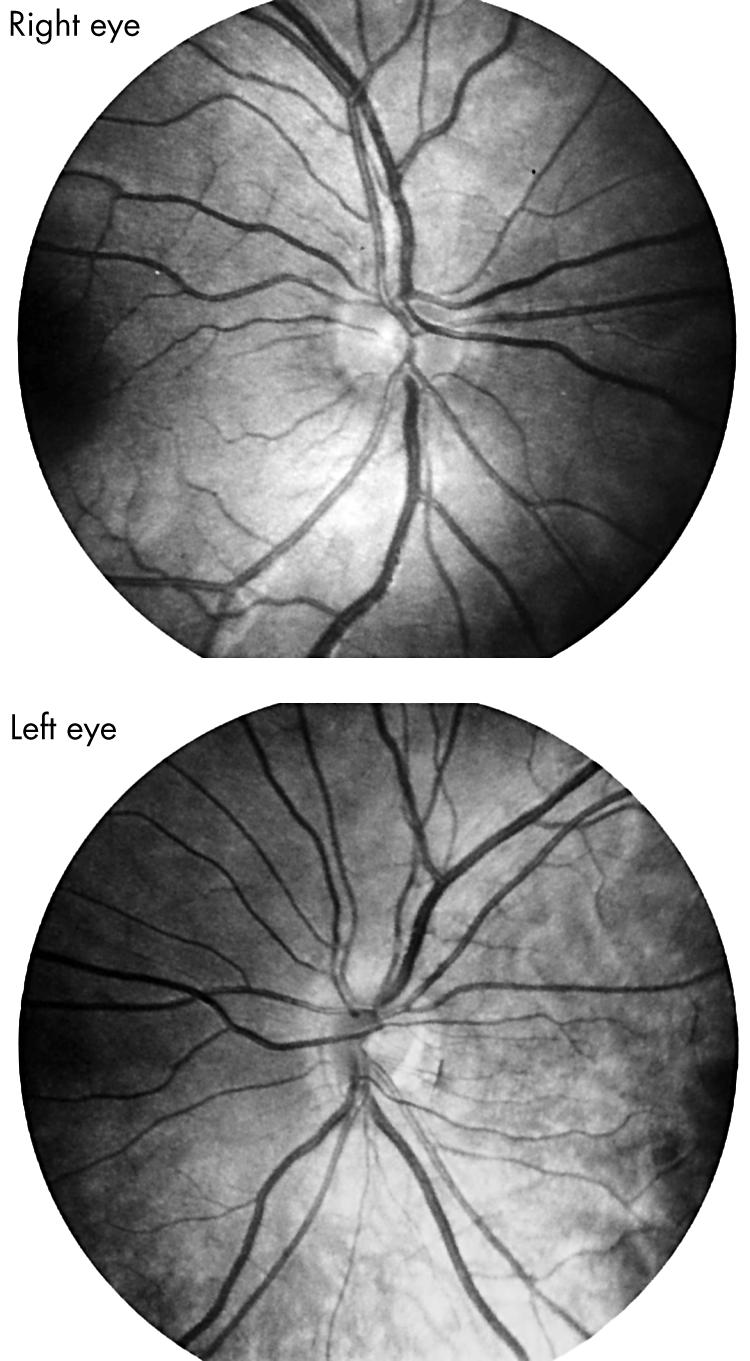
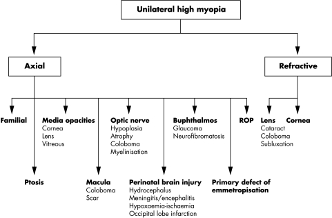
Methods: Medical records of 48 children (aged 4 months to 17 years; mean age 6.8 years) with unilateral high myopia (5 dioptres or more) seen consecutively by the author during a 15 year period were reviewed. 45 (94%) of the 48 patients had unilateral axial myopia.
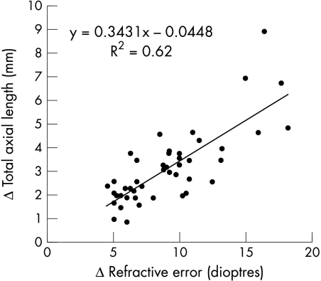
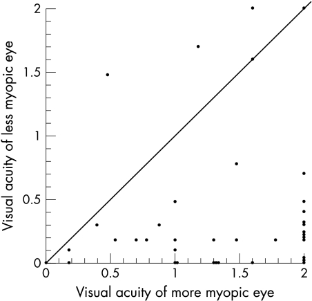
Results: The mean refractive difference between paired eyes was 9.4 (SD 3.6) dioptres and the more myopic eye was on average 3.3 (1.8) mm longer than the less myopic eye. All but three of the patients had an ocular disorder associated with reduced acuity, central nervous system abnormality, or family history of high myopia.
Conclusion: Clinical conditions associated with unilateral high myopia can be identified in the majority of patients and often account for the associated visual impairment.
Figures
References
-
- Sperduto RD, Siegel D, Roberts J, et al. Prevalence of myopia in the United States. Arch Ophthalmol 1983;101:405–7. - PubMed
-
- Angle J, Wissman DA. The epidemiology of myopia. Am J Epidemiol 1980;111:220–8. - PubMed
-
- Curtin BJ, ed. Anisometropic myopia. In: The myopias: basic science and clinical management. Philadelphia: Harper and Row, 1985;chapter 17:449–54.
-
- Sorsby A, Leary GA, Richards MJ. The optical components in anisometropia. Vis Res 1962;2:43–51.
-
- Pollard ZF, Manley D. Long-term results in the treatment of unilateral high myopia with amblyopia. Am J Ophthalmol 1974;78:397–9. - PubMed
Publication types
MeSH terms
LinkOut - more resources
Full Text Sources
Other Literature Sources