. 2003 Aug 1;22(15):3772-82.
doi: 10.1093/emboj/cdg382.

The guanylate binding protein-1 GTPase controls the invasive and angiogenic capability of endothelial cells through inhibition of MMP-1 expression

Affiliations

The guanylate binding protein-1 GTPase controls the invasive and angiogenic capability of endothelial cells through inhibition of MMP-1 expression

Eric Guenzi et al. EMBO J. .

Abstract

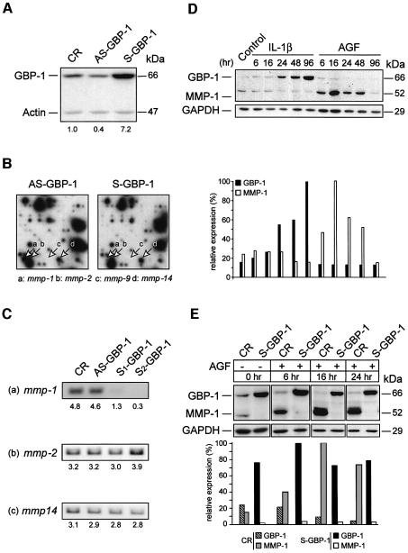
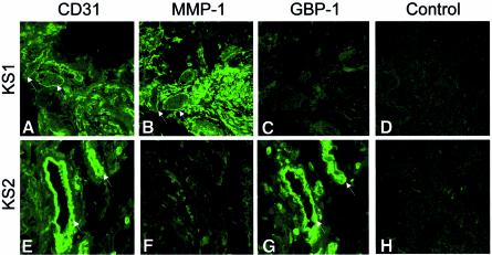
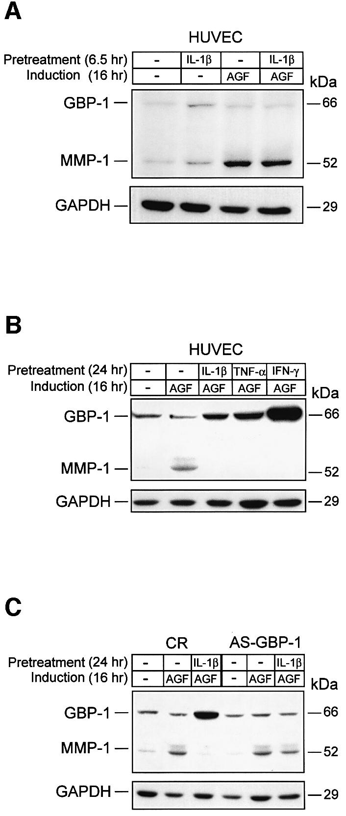
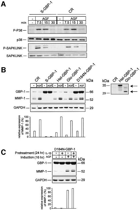
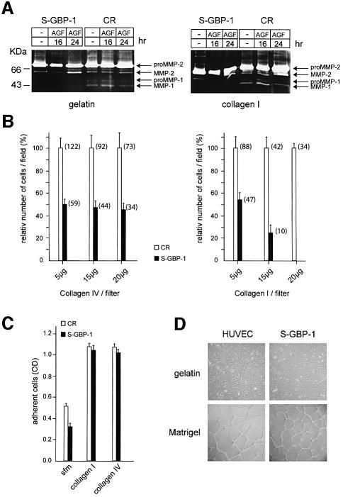
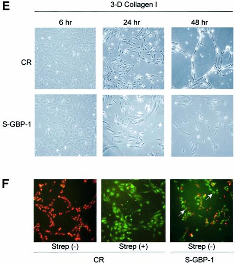
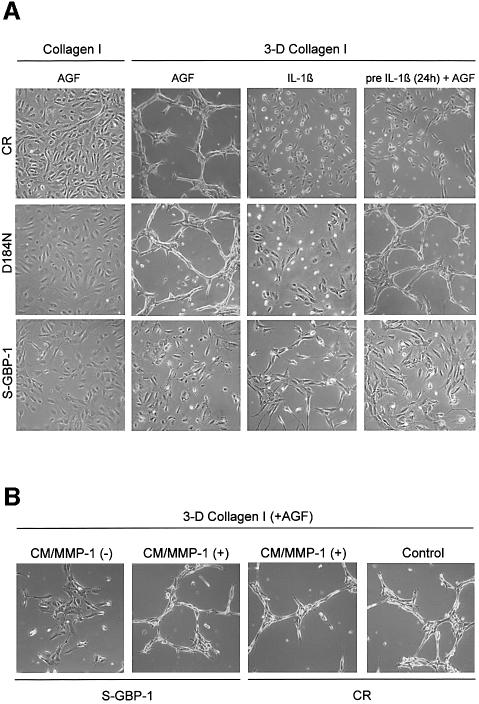
Expression of the large GTPase guanylate binding protein-1 (GBP-1) is induced by inflammatory cytokines (ICs) in endothelial cells (ECs), and the helical domain of the molecule mediates the repression of EC proliferation by ICs. Here we show that the expression of GBP-1 and of the matrix metalloproteinase-1 (MMP-1) are inversely related in vitro and in vivo, and that GBP-1 selectively inhibits the expression of MMP-1 in ECs, but not the expression of other proteases. The GTPase activity of GBP-1 was necessary for this effect, which inhibited invasiveness and tube-forming capability of ECs in three-dimensional collagen-I matrices. A GTPase-deficient mutant (D184N-GBP-1) operated as a transdominant inhibitor of wild-type GBP-1 and rescued MMP-1 expression in the presence of ICs. Expression of D184N-GBP-1, as well as paracrine supplementation of MMP-1, restored the tube-forming capability of ECs in the presence of wild-type GBP-1. The latter finding indicated that the inhibition of capillary formation is specifically due to the repression of MMP-1 expression by GBP-1, and is not affected by the anti-proliferative activity of the helical domain of GBP-1. These findings substantiate the role of GBP-1 as a major regulator of the anti-angiogenic response of ECs to ICs.


Figures

None
Fig. 1. GBP-1 expression is inversely related with matrix metalloproteinase-1 (MMP-1) expression in endothelial cells. (A) Western blot analysis of GBP-1 expression in CR-, AS-GBP-1- and S-GBP-1-transduced HUVECs using a polyclonal rabbit anti-GBP-1 antibody. Corresponding signal intensities were densitometrically determined and normalized to actin in each lane. The relative increase of GBP-1 expression compared with CR-transduced HUVECs is given below. (B) Gene expression profile of AS-GBP-1- and S-GBP-1-transduced HUVECs using Atlas™ Human 1.2 array I. Signals corresponding to the main matrix metalloproteinases expressed in endothelial cells are indicated by arrows: a, mmp-1; b, mmp-2; c, mmp-9; d, mmp-14. (C) RT–PCR analysis of mmp-1, mmp-2 and mmp-14 expression in CR-, AS-GBP-1- and two independent S-GBP-1-transduced (S1, S2) HUVEC cultures. Corresponding signal intensities were densitometrically determined (arbitrary values) and are given below. (D) Western blot analysis of GBP-1 and MMP-1 expression in HUVECs either unstimulated (control) or stimulated with AGF (VEGF and bFGF, 10 ng/ml each) or IL-1β (200 U/ml) for the indicated periods of time. Corresponding signal intensities were densitometrically determined and normalized to GAPDH for each lane (lower panel). (E) Western blot analyses of GBP-1 and MMP-1 expression in CR- and S-GBP-1-transduced HUVECs incubated for the indicated periods of time with (+) or without (–) AGF (10 ng/ml). Corresponding signal intensities were densitometrically determined and normalized to GAPDH for each lane (lower panel). The monoclonal rat anti-GBP-1 antibody (mAb 1B1) was used for the detection of GBP-1 in (D) and (E).
None
Fig. 2. GBP-1 expression is inversely related to MMP-1 expression in endothelial cells in Kaposi’s sarcoma (KS). (AC and EG) Immunofluorescence staining of consecutive sections of two different KS biopsies (KS1, KS2) for the detection of the endothelial cell-associated antigen CD31, of MMP-1 and of GBP-1. Corresponding CD31- and MMP-1-positive, and CD31- and GBP-1-positive vessel endothelial cells are labeled with an arrow. MMP-1 (D) and GBP-1 (H) expressing KS sections subjected to the staining procedure without the anti-MMP-1 (D) and the anti-GBP-1 (H) antibody, respectively.
None
Fig. 3. GBP-1 mediates the IC-induced inhibition of MMP-1 expression in vitro. Western blot analyses of GBP-1 and MMP-1 expression in HUVECs. (A) Cells were pre-treated without (–) or with IL-1β (6.5 h, 200 U/ml) and subsequently incubated without (–) or with AGF (VEGF and bFGF, 10 ng/ml each) for 16 h. (B) Cells were pre-treated without (–) or with IL-1β (200 U/ml), TNF-α (300 U/ml) or IFN-γ (100 U/ml) for 24 h, and subsequently stimulated with AGF (10 ng/ml) for 16 h. (C) CR- and AS-GBP-1-transduced HUVECs were pre-treated without (–) or with IL-1β (24 h, 200 U/ml), and subsequently stimulated with AGF (10 ng/ml) for 16 h. Immunochemical detection of GAPDH indicates, for each lane, the amount of protein blotted onto the membranes.
None
Fig. 4. The GTPase activity of GBP-1 is necessary to inhibit MMP-1 expression. (A) Western blot analysis of phospho-P38 (P-P38) and phospho-SAPK/JNK (P-SAPK/JNK) MAPK expression in S- and CR-GBP-1-transduced HUVECs stimulated with AGF (VEGF and bFGF, 10 ng/ml each), except control (–), for the indicated periods of time. (B) Left panel, western blot analyses of GBP-1 and MMP-1 expression in CR-, S-GBP-1-, Hel-GBP-1-, Glo-GBP-1- and D184N-GBP-1-transduced HUVECs stimulated with AGF (10 ng/ml) for 16 h (upper panel). Corresponding signal intensities were densitometrically determined and normalized to GAPDH in each lane (lower panel). Right panel, western blot analyses of the expression of the C-terminal helical domain (Hel-GBP-1, upper arrow) and the N-terminal globular domain (Glo-GBP-1, lower arrow) in HUVECs transduced with the respective retroviral vectors. CR-transduced HUVECs were used as a negative control (CR). A polyclonal rabbit anti-GBP-1 antibody was used. (C) Western blot analyses of GBP-1 and MMP-1 expression in D184N-GBP-1-transduced HUVECs (upper panel). Cells were pre-treated without (–) or with IL-1β (24 h, 200 U/ml), and subsequently incubated without (–) or with AGF (10 ng/ml) for 16 h. The rat monoclonal anti-GBP-1 (mAb, 1B1) antibody was used for detection of GBP-1. Immunochemical detection of GAPDH demonstrates that equal concentrations of protein were blotted onto the membranes (middle panel). Corresponding signal intensities were densitometrically determined and normalized to GAPDH in each lane (lower panel).
None
Fig. 5. GBP-1 inhibits migration and tube formation of HUVECs on collagen I matrices. (A) Zymography analysis using gelatin (left panel) or collagen I (right panel) as substrates. Cell culture supernatants of S-GBP-1- and CR-transduced HUVECs stimulated with AGF (VEGF and bFGF, 10 ng/ml each), except control (–), were used for the times indicated. The bands corresponding to the pro- and active forms of MMP-1 and MMP-2 are indicated (arrows). (B) Invasion assay of CR- and S-GBP-1-transduced HUVECs in collagen IV (left panel) and collagen I (right panel). The mean values obtained in three different experiments are shown [± standard deviations (SDs)]. The numbers of transmigrated CR-transduced cells in the respective experiments were set to 100% (white columns), and the relative numbers of S-GBP-1-transduced cells (black columns) were calculated accordingly. The absolute numbers of transmigrated cells are given in parentheses. (C) Adhesion assay of CR- and S-GBP-1-transduced HUVECs to collagen I and collagen IV matrices. The mean values obtained in six independent experiments are shown (± SD). Serum-free medium (sfm) without collagen was used as a control. (D) Phase contrast pictures of HUVECs or S-GBP-1-transduced HUVECs cultured for 24 h on gelatin (upper panel) or Matrigel (lower panel) in the presence of AGF (10 ng/ml). (E) Tube formation of CR- or S-GBP1-transduced HUVECs in 3D collagen I matrices in the presence of AGF (10 ng/ml) and PMA (0.1 µM). (F) In situ detection of apoptosis of CR- and S-GBP1-transduced HUVECs in 3D collagen I matrices after 24 h treated as in (E). Apoptosis was induced by treatment with streptonigrin (Strep, 1 µM) for 3 h. Non-apoptotic, CR-transduced HUVECs are stained red [CR, Strep(–)]. Apoptotic cells are stained green [Strep(+)]. White arrows indicate apoptotic cells (green) in the tube formation assay with S-GBP-1-transduced HUVECs.
None
Fig. 6. Tube formation in 3D collagen I matrices requires MMP-1 expression and is independent of cell proliferation. (A) Left panel, morphology of CR-, D184N- and S-GBP-1-transduced HUVECs cultivated on collagen I for 48 h in the presence of AGF (10 ng/ml). Middle two panels, tube formation of transduced HUVECs in 3D collagen I matrices in the presence of AGF (10 ng/ml) or IL-1β (200 U/ml) for 48 h. Right panels, tube formation of the respective transduced cells pre-treated for 24 h with IL-1β (200 U/ml) and subsequently grown in 3D collagen I matrices for 48 h. (B) Tube formation of S-GBP1- and CR-transduced HUVECs in 3D collagen I matrices. Culture medium (CM) of HUVECs stimulated with AGF for 16 h [CM/MMP-1(+)] or of unstimulated HUVECs [CM/MMP-1(–)] was added to AGF- (10 ng/ml) treated CR- and S-GBP1-transduced HUVECs in collagen I matrices for 48 h. Right, CR-transduced HUVECs in 3D collagen I matrices in the presence of AGF (control).

Similar articles

Cited by

References

    1. Benbow U., Tower,G.B., Wyatt,C.A., Buttice,G. and Brinckerhoff,C.E. (2002) High levels of MMP-1 expression in the absence of the 2G single nucleotide polymorphism is mediated by p38 and ERK1/2 mitogen-activated protein kinases in VMM5 melanoma cells. J. Cell. Biochem., 86, 307–319. - PubMed
    1. Cardozo A.K., Heimberg,H., Heremans,Y., Leeman,R., Kutlu,B., Kruhoffer,M., Orntoft,T. and Eizirik,D.L. (2001) A comprehensive analysis of cytokine-induced and nuclear factor-κB-dependent genes in primary rat pancreatic beta-cells. J. Biol. Chem., 276, 48879–48886. - PubMed
    1. Carmeliet P. and Jain,R.K. (2000) Angiogenesis in cancer and other diseases. Nature, 407, 249–257. - PubMed
    1. Cheng Y.S., Colonno,R.J. and Yin,F.H. (1983) Interferon induction of fibroblast proteins with guanylate binding activity. J. Biol. Chem., 258, 7746–7750. - PubMed
    1. Cheng Y.S., Patterson,C.E. and Staeheli,P. (1991) Interferon-induced guanylate-binding proteins lack an N(T)KXD consensus motif and bind GMP in addition to GDP and GTP. Mol. Cell. Biol., 11, 4717–4725. - PMC - PubMed

Publication types

MeSH terms