. 2003 Aug 1;22(15):3971-82.
doi: 10.1093/emboj/cdg369.

The apolipoprotein B mRNA editing complex performs a multifunctional cycle and suppresses nonsense-mediated decay

Affiliations

The apolipoprotein B mRNA editing complex performs a multifunctional cycle and suppresses nonsense-mediated decay

Ann Chester et al. EMBO J. .

Abstract

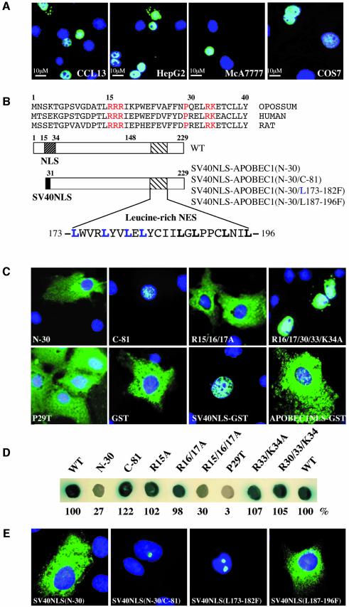
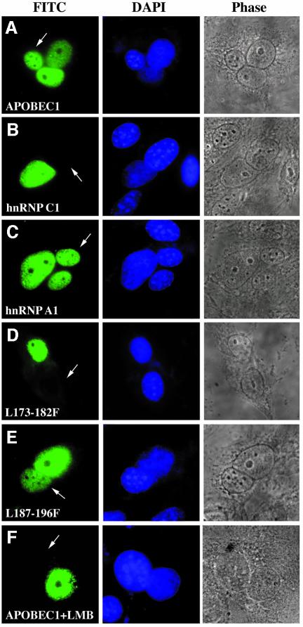
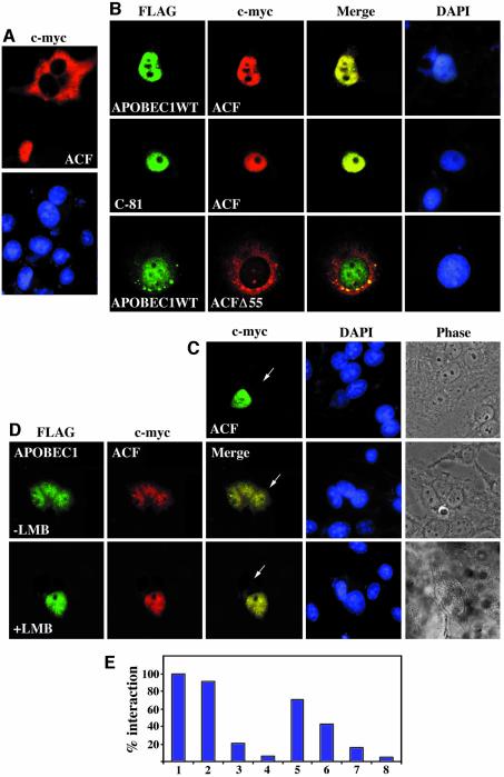
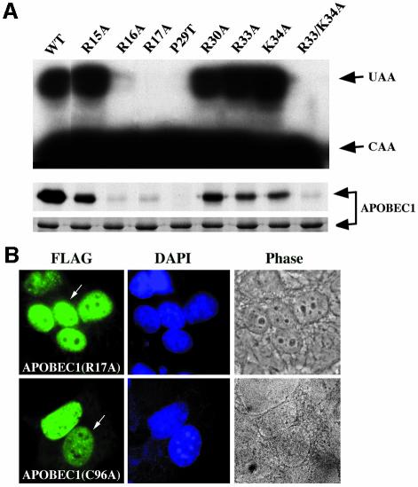
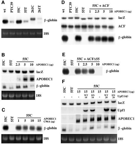
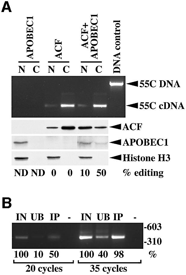
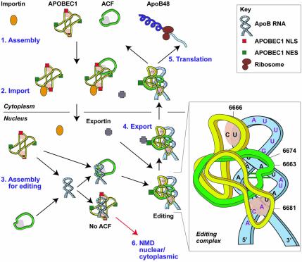
The C to U editing of apolipoprotein B (apoB) mRNA is mediated by a minimal complex composed of an RNA-binding cytidine deaminase (APOBEC1) and a complementing specificity factor (ACF). This editing generates a premature termination codon and a truncated open reading frame. We demonstrate that the APOBEC1-ACF holoenzyme mediates a multifunctional cycle. The atypical APOBEC1 nuclear localization signal is involved in RNA binding and is used to import ACF into the nucleus as cargo. APOBEC1 alone induces nonsense-mediated decay (NMD). The APOBEC1-ACF complex edits and remains associated with the edited RNA to protect it from NMD. The APOBEC1 nuclear export signal is involved in the export of ACF and the edited apoB mRNA together, to the site of translation.


Figures

None
Fig. 1. APOBEC1 has a minimal bipartite N-terminal NLS and a leucine-rich NES. (A) Nuclear localization of C-terminal FLAG-tagged APOBEC1 in transiently transfected CCL13, HepG2, McA777 and COS-7 cells. The images show FITC overlaid with DAPI-stained nuclei. (B) Alignment of the N-terminal sequence of APOBEC1. The amino acid residues in the atypical bipartite NLS are shown in red and on a diagram of APOBEC1 (dark-hatched box) together with the leucine-rich NES (light-hatched box). The N-terminal 30 amino acids of APOBEC1 replaced with SV40NLS [SV40NLS–APOBEC1(N-30)], deletion of the C-terminal 81 amino acids of SV40NLS–APOBEC1(N-30) [SV40NLS–APOBEC1(N-30/C-81)] and leucines 173–182 or 187–196 mutated to phenylalanine [SV40NLS–APOBEC1(N-30/L173–182F or L187–197F)]. Leucines 173–182 are highlighted in blue, and 187–197 are shown in bold. (C) Intracellular localization of the APOBEC1 deletion (N-terminal deletion N-30, C-terminal deletion C-81) and combined mutants (R15/16/17A, R16/17/30/33/K34A) and point mutant (P29T) in COS-7 cells. Localization of GST, SV40NLS–GST and APOBEC1 NLS (1–44)–GST. The images show protein stained with FITC and overlaid with DAPI-stained nuclei. (D) Interaction of APOBEC1 and its mutants with importin α, analysed in the yeast two-hybrid system. Blue colonies denote interaction and white colonies denote no interaction. The strength of the interactions was also measured by liquid β-galactosidase assays and given as a percentage of the wild-type interaction. (E) Subcellular localization of the FITC images of SV40NLS–APOBEC1(N-30) and its mutant C-81, L173–182F and L187–196F proteins overlaid with DAPI.
None
Fig. 2. Exportin-mediated shuttling of APOBEC1 requires L173–L182. FLAG-tagged APOBEC1 wild-type (APOBEC1WT) (A), hnRNP C1 (B) and A1 (C) leucine mutants APOBEC1(L173–182F) (D) and APOBEC1(L187–196F) (E) were transfected into CCL13 cells and fused to mouse NIH-3T3 cells. (F) Heterokaryon of APOBEC1WT incubated with 50 ng/ml leptomycin B (LMB). Proteins were stained with anti-FLAG antibody and visualized with anti-mouse FITC (left). Nuclei were stained with DAPI (middle). Phase-contrast images of the heterokaryons are shown (right). The arrowhead indicates the mouse nuclei in the mouse–human heterokaryons.
None
Fig. 3. APOBEC1 transports ACF as its cargo to and from the nucleus. (A) Subcellular localization of ACF c-myc (ACF) in CCL13 cells visualized with anti-c-myc-Cy3-conjugated antibody. (B) Subcellular localization of FLAG-tagged APOBEC1 wild-type (APOBEC1WT) or deletion of the C-terminal 81 amino acids of APOBEC1 (C-81), co-transfected with ACF c-myc (ACF) or APOBEC1WT with ACFΔ55 and visualized with anti-FLAG and anti-c-myc antibodies as before. Merged images show co-localization of ACF with APOBEC1WT and C-81 deletion mutant (top/middle), whereas APOBEC1WT with ACFΔ55 did not co-localize (bottom). DAPI-stained nuclei are also shown. (C) Heterokaryon analysis of nuclear ACF c-myc with anti-c-myc antibody and visualized with anti-mouse FITC. Nuclei were stained with DAPI, and a phase-contrast image of the heterokaryon is shown. (D) Co-transfected APOBEC1WT with ACF analysed by heterokaryon assay in the absence (top) and presence (bottom) of LMB. APOBEC1WT and ACF are visualized as before. Merged images show co-localization of both proteins in the mouse cells (arrowhead) in the absence of LMB. (E) Yeast two-hybrid analysis of the interaction of ACF with (1) APOBEC1, and its mutants (2) C-100, (3) N-15, (4) N-30, (5) R15/16A, (6) R15/16/17A, (7) P29T, and (8) vector control. The strength of the interaction measured by β-galactosidase assays is shown as a percentage of the wild-type interaction.
None
Fig. 4. APOBEC1 NLS and NES are involved in RNA binding. (A) RNA editing (top) and RNA UV cross-linking (middle) properties of GST fusion proteins of APOBEC1WT, point mutants R15, 16, 17, 30 33A, P29T and K34A and combined mutant R33/K34A. Edited (UAA), unedited (CAA) (top), RNA UV cross-linked and Coomassie-stained proteins (bottom) are shown. (B) Heterokaryon analysis of FLAG-tagged APOBEC1 mutants (R17A and C96A) visualized as in Figure 3.
None
Fig. 5. ACF protects apoB RNA from APOBEC1-induced NMD. (A) Northern blot analysis of total RNA from CCL13 cells transfected with 10 µg of plasmid DNA containing β-globin wild-type (wt), PTC 39, 55 or 261 nucleotides of unedited (C) and edited (T) apoB RNA inserted in-frame in exon 2 of β-globin wild type (55C, 55T, 261C and 261T) in the same position as PTC 39. Northern blots probed with β-globin cDNA. Ethidium bromide staining of the 18S RNA from the corresponding gel is shown. (B) Northern blot analysis of total RNA obtained from cells transfected with plasmids 55T or 55C (10 µg) together with 5 µg of lacZ and 0–10 µg of APOBEC1 in a total of 30 µg of DNA per transfection and probed with lacZ, APOBEC1 and β-globin cDNAs. In some experiments, empty vector plasmid DNA was used to make up the total concentration of DNA to 30 µg. (C) Northern blot analysis of total RNA as in (B) except APOBEC1 was replaced with APOBEC1(C96A) catalytically inactive mutant and probed with β-globin cDNA. Other probes are not shown. (D) Northern blot analysis of total RNA from cells transfected with β-globin wild-type (wt), PTC 39, 55T or 55C (10 µg) together with 5 µg of lacZ, 30 µg of ACF and 1–10 µg of APOBEC1 in a total of 55 µg of DNA per transfection, and probed with lacZ, ACF and β-globin cDNAs. Empty vector plasmid DNA was used to make up the total concentration of DNA to 55 µg. (E) Northern blot analysis of total RNA as in (D) except ACF was replaced with ACFΔ55 and probed as in (D). Only the β-globin probe is shown. (F) Northern blot analysis of total RNA from cells transfected with 55T or 55C (10 µg) together with 5 µg of lacZ, 15 µg of APOBEC1 and 10–20 µg of Upf1 wild-type (WT) or R844C dominant-negative mutant (DN) in a total of 50 µg of DNA per transfection and probed with lacZ, Upf1, APOBEC1 and β-globin cDNAs. Empty vector plasmid DNA was used to make up the total concentration of DNA to 50 µg.
None
Fig. 6. The APOBEC1–ACF complex contains edited RNA. (A) RT–PCR products (398 bp) derived from immunoprecipitated RNA isolated from nuclear (N) and cytoplasmic (C) extracts obtained by transfecting APOBEC1, ACF or both in CCL13 cells along with the 55C β-globin DNA (55C). Transfected 55C plasmid containing genomic DNA generated a 1390 bp PCR product. Western blot analysis of ACF, APOBEC1 and histone H3 from identical extracts used above and the percentage of apoB RNA editing is shown. (B) RT–PCR (20 and 35 cycles) of input (IN), unbound (UB) and immunoprecipitated (IP) RNA from cytoplasmic extracts containing APOBEC1–ACF. The amount of product is shown as a percentage of the input RNA-derived product for each experimental condition.
None
Fig. 7. A model for the assembly, editing and transport of the apoB mRNA editing complex. (1) The initial cytoplasmic event of apoB mRNA editing is the heterotrimerization of ACF with an APOBEC1 dimer. (2) Interaction of importin α with APOBEC1 NLS targets the APOBEC1–ACF complex to the nucleus with ACF as cargo. The importin complex is shown as a single entity consisting of importin α and β subunits. (3) In the nucleus, this complex dissociates or relaxes. Assembly of the editing complex on the apoB RNA occurs via the site-specific binding of ACF to the RNA and the recruitment or repositioning of APOBEC1. C6666 is edited to U, generating a PTC. The editing complex remains associated with the RNA and protects it from NMD. (4) Conformational changes that take place during the assembly of the editing complex expose the APOBEC1 NES. Exportin 1 mediates the export of the apoB RNP complex to the cytoplasm. (5) Translation of edited apoB mRNA generates apoB48. (6) In the absence of ACF, non-specific binding of APOBEC1 to the RNA generates multiple PTCs. RNA with PTC undergoes nuclear or cytoplasmic NMD. The insert shows a schematic representation of the editing complex; unpaired nucleotides are shown in red, APOBEC1 and ACF binding sites are shaded.

Similar articles

Cited by

References

    1. Blanc V., Navaratnam,N., Henderson,J.O., Anant,S., Kennedy,S., Jarmuz,A., Scott,J. and Davidson,N.O. (2001a) Identification of GRY-RBP as an apolipoprotein B RNA-binding protein that interacts with both apobec-1 and apobec-1 complementation factor to modulate C to U editing. J. Biol. Chem., 276, 10272–10283. - PubMed
    1. Blanc V., Henderson,J.O., Kennedy,S. and Davidson,N.O. (2001b) Mutagenesis of apobec-1 complementation factor reveals distinct domains that modulate RNA binding, protein–protein interaction with apobec-1 and complementation of C to U RNA-editing activity. J. Biol. Chem., 276, 46386–46393. - PubMed
    1. Chester A., Scott,J., Anant,S. and Navaratnam,N. (2000) RNA editing: cytidine to uridine conversion in apolipoprotein B mRNA. Biochim. Biophys. Acta, 1494, 1–13. - PubMed
    1. Conti E., Uy,M., Leighton,L., Blobel,G. and Kuriyan,J. (1998) Crystallographic analysis of the recognition of a nuclear localization signal by the nuclear import factor karyopherin α. Cell, 94, 193–204. - PubMed
    1. Dingwall C. and Laskey,R. (1991) Nuclear targeting sequences—a consensus? Trends Biochem. Sci., 16, 478–481. - PubMed

Publication types