. 2003 Aug 1;22(15):3992-4002.
doi: 10.1093/emboj/cdg387.

Permanent cell cycle exit in G2 phase after DNA damage in normal human fibroblasts

Affiliations

Permanent cell cycle exit in G2 phase after DNA damage in normal human fibroblasts

Fabienne Baus et al. EMBO J. .

Abstract

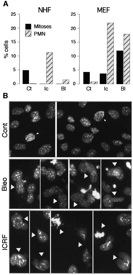
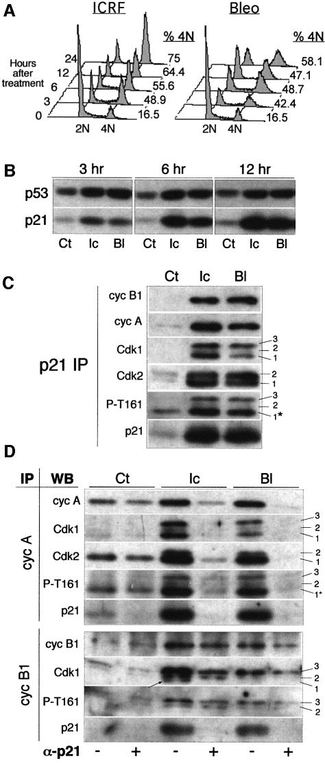
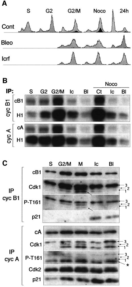
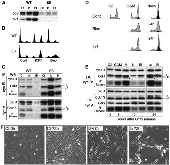
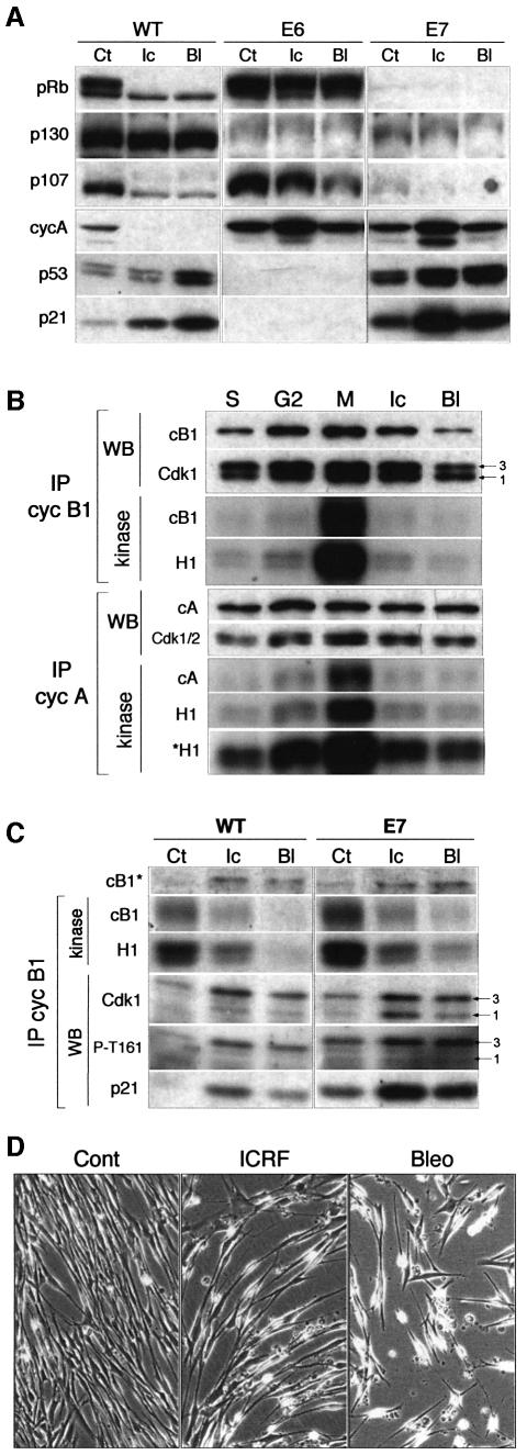
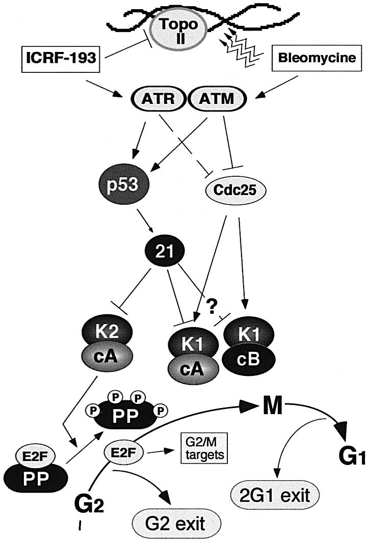
Although the Cdk inhibitor p21(Waf1/Cip1), one of the transcriptional targets of p53, has been implicated in the maintenance of G(2) arrest after DNA damage, its function at this stage of the cell cycle is not really understood. Here, we show that the exposure of normal human fibroblasts (NHFs) to genotoxic agents provokes permanent cell cycle exit in G(2) phase, whereas mouse embryo fibroblasts and transformed human cells progress through mitosis and arrest in G(1) without intervening cytokinesis. p21(Waf1/Cip1) exerts a key role in driving this G(2) exit both by inhibiting cyclin B1-Cdk1 and cyclin A-Cdk1/2 complexes, which control G(2)/M progression, and by blocking the phosphorylation of pRb family proteins. NHFs with compromised pRb proteins could still efficiently arrest in G(2) but were unable to exit the cell cycle, resulting in cell death. Our experiments show that, when under continuous genotoxic stress, normal cells can reverse their commitment to mitotic progression due to passage through the restriction point and that mechanisms involving p21(Waf1/Cip1) and pocket proteins can induce exit in G(2) and G(1).


Figures

None
Fig. 1. Mouse embryo fibroblasts (MEFs) have a non-functional G2/M checkpoint in comparison with normal human fibroblasts (NHFs). (A) Percentage of cells in mitosis or with aberrant post-mitotic nuclei (PMN) in untreated (Ct) and 24 h ICRF-193-treated (Ic) and bleomycin-treated (Bl) NHF and MEF cultures. For each situation, at least 500 cells were scored. (B) Micrographs (Hoechst) showing untreated MEFs (Cont) and some characteristic bleomycin- and ICRF-193-induced mitotic and aberrant PMN (arrowheads) after 24 h exposure. Arrows point to normal mitoses in untreated cells.
None
Fig. 2. In response to DNA damage, p21 targets Cdks regulating the G2/M transition. (A) Cell cycle profiles of exponentially growing cells exposed to ICRF-193 and bleomycin for the indicated times. Percentage of cells containing 4N DNA content is shown on the right of each FACS profile. (B) p53 and p21 protein levels in untreated cells (Ct) or cells exposed to ICRF-193 (Ic) or bleomycin (Bl) for the indicated times. (C) Western blot analysis of p21 immunoprecipitates (IPs) isolated from control cells and those treated for 12 h with ICRF-193 or bleomycin. Cdk1 migrates as three bands (1–3), whereas Cdk2 migrates as two bands (1 and 2). Number 1 points to CAK-phosphorylated/Cdc25-dephosphorylated Cdk isoforms (potentially active). Note that antibody directed against phospho-Thr161 (P-T161) recognizes both Cdk1 and Cdk2 (asterisk). (D) Immunoblot analysis of cyclin complexes isolated before and after p21 immunodepletion. Cell lysates prepared from untreated cells and those treated for 12 h (as above) were immunodepleted for p21-associated complexes by successive incubations with an anti-p21 antibody (+). Mock samples of the same extracts were incubated with protein A beads alone (-). Cyclin IPs were examined for cyclin, Cdk and p21 content. Numbers 1–3 point to the different Cdk1 and Cdk2 isoforms. In cyclin B1 IP, the arrow points to the hypophosphorylated Cdk1 (1) isoform. Note that antibody directed against P-T161 recognizes both Cdk1 and Cdk2 (asterisk).
None
Fig. 3. Bleomycin and ICRF-193 block mitosis by preventing activation of mitotic cyclin–Cdk complexes. (A) Flow cytometry analysis of normal human fibroblasts synchronized at the G1/S transition and released into the cell cycle in the absence or presence of bleomycin and ICRF-193. Mitotic cells were labelled by using the antibody directed against phospho-histone H3 (black-shaded profile). Untreated cells were harvested and analysed at 3 h (S phase), 7 h (G2 phase) and 9.5 h (G2/M transition) after release from the G1/S block. ICRF-193- and bleomycin-treated samples were collected at the time when most control cells have entered mitosis (G2/M), 2 h later in the presence of nocodazole (Noco), and after 24 h. (B) Histone H1 kinase (H1) activity and cyclin autophosphorylation of cyclin B1 (cB1) and cyclin A (cA) immunoprecipitates (IPs) isolated from corresponding cell extracts. Noco denotes the cultures to which nocodazole was added (2 h) to ‘trap’ the cells in metaphase. (C) Western blot analysis of cyclin B1 and cyclin A IPs isolated from synchronized untreated and drug-treated cells showing cyclin, Cdk and p21 content. In cyclin B1 immunoblots, arrows point to the hypophosphorylated cyclin B1-associated Cdk1 band that specifically accumulates in mitotis. In drug-treated cells, the Cdk1 band migrating at the same position, but not recognized by an antibody directed against phospho-Thr161 (P-T161), is removed by p21 immunodepletion (cf. Figures 2D and 6C). Note that the lowest band (asterisk) in cyclin A IP is composed of hyperphosphorylated Cdk2 and hypophosphorylated Cdk1 isoforms, as anti-P-T161 antibody recognizes both Cdks. Numbered arrows point to the different Cdk isoforms.
None
Fig. 4. Impaired but differential response to ICRF-193 and bleomycin in normal human fibroblasts (NHFs) deficient for p53/p21 pathway. (A) p53 and p21 proteins levels in untreated (Ct), ICRF-193-treated (Ic) and bleomycin-treated (Bl) wild-type (WT) and E6 NHFs. Note that the low level of p53 detected in E6 cells did not increase after drug treatment. (B) Cell cycle profiles of WT and E6 NHFs exposed to ICRF-193 or bleomycin for 24 h. (C) Histone H1 kinase activity and western blot (WB) analysis of cyclin B1 and cyclin A immunoprecipitates (IPs) isolated from untreated (Ct), ICRF-193- and bleomycin-treated WT and E6 cells. Numbers 1–3 point to the different Cdk isoforms. (D) Flow cytometry analysis (FACS) of E6 cells synchronized at G1/S boundary and released into the cell cycle in the absence (Cont) or presence of the indicated drug. Mitotic cells were labelled by using the antibody directed against phospho-histone H3 (black-shaded profile). Untreated cells were harvested at 6 h (G2 phase) and 8 h (G2/M transition) and in the presence of nocodazole (8 + 2 h). Drug-treated cells were harvested 10 and 24 h after release from the block. (E) Histone H1 kinase activity and WB analysis of cyclin B1 and cyclin A IPs isolated from synchronized untreated (Ct) and ICRF-193- and bleomycin-treated E6 cells. Extracts were analysed from the cells harvested at the indicated times after release from the G1/S block (cf. the FACS analysis above). Arrows point to the hypophosphorylated cyclin B1-associated Cdk1 (isoform 1), which is absent in S phase and enriched in mitotic cells. In the cyclin A IPs, the lowest PSTAIRE-recognized band (1) consists of both hypophosphorylated Cdk1 and CAK-phosphorylated Cdk2 isoforms. Numbers 1–3 point to the different Cdk isoforms. (F) Micrographs taken from a video-microscopy sequence showing E6 cells in the absence (Ct) of the drugs and those exposed for 72 h to bleomycin or ICRF-193. Untreated cells were shown at the beginning of the experiment (0 h; at the time when the drugs were added) and after 72 h. Note the dying cells in drug-treated cultures.
None
Fig. 5. DNA damage leads to irreversible cell cycle exit in G2. (A) Western blot analysis of protein lysates prepared from exponentially growing normal human fibroblasts (NHFs) untreated (Ct) or exposed at various times to ICRF-193 (Ic) and bleomycin (Bl). G0 denotes contact inhibited NHFs. To assess different aspects of regulation contributing to cell cycle arrest and exit, levels of pocket proteins (pRb, p130 and p107), Cdk inhibitors p21 and p27, and cyclin A were monitored. Note the rapid accumulation of hypophosphorylated forms of pocket proteins (arrows) while cells still contained high levels of cyclin A. (B) Western blot analysis showing phosphorylation status of pocket proteins in control and drug-treated synchronized NHFs. FACS profile of cells exposed to ICRF-193 and bleomycin collected 24 h after the G1/S block is shown in Figure 3A. Biochemical analysis of corresponding cyclin immunoprecipitates is shown in Figure 3A and B. (C) Western blot analysis showing pRb and mitotic cyclin levels in control and drug-treated synchronized E6 cells. The same samples are shown in Figure 4. Chk2 status was monitored to assess efficient signalling in response to DNA damage in E6 cells. (D) Cell size analysis (flow cytometry) of control G2 cells versus G2 cells that were exposed to ICRF-193 or bleomycin for 7 and 24 h. (E) Micrographs showing cell morphology and β-gal staining of untreated NHFs (a) and cultures exposed to ICRF-193 for 24 h (b) and 72 h (c) after treatment. As positive controls, NHFs expressing oncogenic ras (d) and senescent culture (e) are shown.
None
Fig. 6. Active pocket proteins are required for cell cycle exit in G2 after genotoxic stress. (A) Western blot analysis of protein extracts prepared from exponentially growing wild-type (WT) and E6- and E7-expressing normal human fibroblasts (NHFs) that were untreated (Ct) or exposed to ICRF-193 (Ic) and bleomycin (Bl) for 24 h. Note the persistent high levels of cyclin A in drug-treated E6 and E7 cells. (B) Histone H1 kinase activity and western blot (WB) analysis of cyclin B1 and cyclin A immunoprecipitates (IPs) isolated from synchronized untreated, ICRF-193- and bleomycin-treated E7 fibroblasts. Untreated cells were harvested in S, G2 and M phases. Nocodazole was added to both control and treated cells to ‘trap’ mitotic cells. Autoradiographs show both histone H1 and autophosphorylated cyclins. To accentuate the increase cyclin A-associated kinase activity in mitosis, both short and long (*H1) autoradiograph exposure are shown. The same immunoblots were analysed for the presence of cyclins (A and B1) and Cdk. Numbered arrows point to the different Cdk1 phospho- isoforms. (C) Kinase activity and WB analysis of cyclin B1 IPs isolated from untreated (Ct), ICRF-193- and bleomycin-treated (6 h) asynchronously growing WT and E7 cells. Numbered arrows point to the hyperphosphorylated (3) and hypophosphorylated (1) cyclin B1-associated Cdk1 isoforms. Note that the latter isoform does not appear to be phosphorylated at T161 in G2-arrested cells. (D) Prolonged exposure to ICRF-193 and bleomycin provokes cell death in spite of efficient G2 arrest. The micrographs (extracted from a video-microscopy sequence) show control and ICRF-193- and bleomycin-treated cultures after 48 h.
None
Fig. 7. Role of p21 in directing cell cycle exit after DNA damage in G2. In response to DNA damage (bleomycin) and the inhibition of topoisomerase II (ICRF-193), ATM and ATR kinases, respectively, are activated (Deming et al., 2001). In E6 cells, the inactivation of Cdc25 via the ATR pathway by ICRF-193 is less efficient. Cell cycle arrest is accomplished by a network of enzymes that block activation of the mitotic kinases cyclin A–Cdk1 and cyclin B1–Cdk1. The question mark refers to the fact that the role of p21 in inactivating cyclin B1–Cdk1 complexes is less well established. In addition to blocking activation of mitotic kinases, p21 also inactivates Cdks involved in the hyperphosphorylation of pocket proteins (PP) of the retinoblastoma family (pRb, p107 and p130) leading to the accumulation of hypophosphorylated (active) pocket proteins. The resulting sequestration of transcription factors of the E2F family would block transcription of a number of genes involved in G2/M progression (Taylor et al., 2001; Ren et al., 2002), thereby driving cell cycle exit in G2. In mouse fibroblasts and tumour-derived cell lines, the exit is accomplished only after mitosis but before cytokinesis, giving rise to tetraploid cells (2G1 exit; cf. Borel et al., 2002). The model also shows that, in the case of ICRF-193-induced G2 arrest, the inactivation of Cdc25 pathways is less efficient than that provoked by double-strand DNA breaks (bleomycin).

Similar articles

Cited by

References

    1. Abraham R.T. (2001) Cell cycle checkpoint signaling through the ATM and ATR kinases. Genes Dev., 15, 2177–2196. - PubMed
    1. Agarwal M.L., Agarwal,A., Taylor,W.R. and Stark,G.R. (1995) p53 controls both the G2/M and the G1 cell cycle checkpoints and mediates reversible growth arrest in human fibroblasts. Proc. Natl Acad. Sci. USA, 92, 8493–8497. - PMC - PubMed
    1. Andreassen P.R., Lacroix,F.B., Lohez,O.D. and Margolis,R.L. (2001a) Neither p21(WAF1) nor 14-3-3σ prevents G2 progression to mitotic catastrophe in human colon carcinoma cells after DNA damage, but p21WAF1 induces stable G1 arrest in resulting tetraploid cells. Cancer Res., 61, 7660–7668. - PubMed
    1. Andreassen P.R., Lohez,O.D., Lacroix,F.B. and Margolis,R.L. (2001b) Tetraploid state induces p53-dependent arrest of nontransformed mammalian cells in G1. Mol. Biol. Cell, 12, 1315–1328. - PMC - PubMed
    1. Borel F., Lohez,O.D., Lacroix,F.B. and Margolis,R.L. (2002) Multiple centrosomes arise from tetraploidy checkpoint failure and mitotic centrosome clusters in p53 and RB pocket protein-compromised cells. Proc. Natl Acad. Sci. USA, 99, 9819–9824. - PMC - PubMed

Publication types