. 2003 Aug 5;100(16):9578-83.
doi: 10.1073/pnas.1133382100. Epub 2003 Jul 24.

Stimulation of NeuroD activity by huntingtin and huntingtin-associated proteins HAP1 and MLK2

Affiliations

Stimulation of NeuroD activity by huntingtin and huntingtin-associated proteins HAP1 and MLK2

Edoardo Marcora et al. Proc Natl Acad Sci U S A. .

Abstract

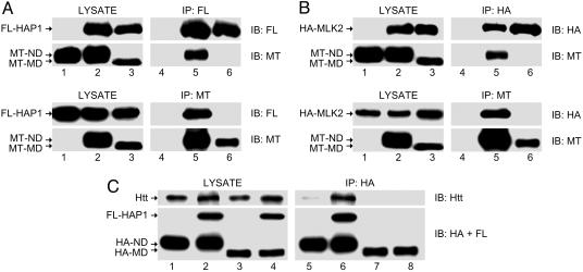
NeuroD (ND) is a basic helix-loop-helix transcription factor important for neuronal development and survival. By using a yeast two-hybrid screen, we identified two proteins that interact with ND, huntingtin-associated protein 1 (HAP1) and mixed-lineage kinase 2 (MLK2), both of which are known to interact with huntingtin (Htt). Htt is a ubiquitous protein important for neuronal transcription, development, and survival, and loss of its function has been implicated in the pathogenesis of Huntington's disease, a neurodegenerative disorder. However, the mechanism by which Htt exerts its neuron-specific function at the molecular level is unknown. Here we report that Htt interacts with ND via HAP1, and that MLK2 phosphorylates and stimulates the activity of ND. Furthermore, we show that Htt and HAP1 facilitate the activation of ND by MLK2. To our knowledge, ND is the first example of a neuron-specific transcription factor involved in neuronal development and survival whose activity is modulated by Htt. We propose that Htt, together with HAP1, may function as a scaffold for the activation of ND by MLK2.


Figures

Fig. 1.
Fig. 1.
ND interacts with HAP1 and MLK2 in the yeast two-hybrid system. To map the domains of interaction among ND, HAP1, and MLK2, the following deletion and point mutants were tested for interaction in the yeast two-hybrid system: ND (amino acids 73–232); ND-ΔLZ (Δ amino acids 169–190); ND-N-term (amino acids 73–97); ND-ΔC-term (amino acids 73–167); ND-bHLH (amino acids 102–154); MyoD-bHLH (mouse MyoD, amino acids 110–160); ND-ΔbHLH (Δ amino acids 102–154); mouse HAP1-A (HAP1, amino acids 247–446); HAP1ΔLZ (Δ amino acids 270–291); mouse MLK2 (corresponding to amino acids 390–460 of human MLK2); MLK2* (L426P). ++, 0–30 min β-galactosidase (β-gal) signal detection time; -, no detectable β-gal signal after 3 h.
Fig. 2.
Fig. 2.
ND interacts with Htt, HAP1, and MLK2 in a neuronal cellular context. (A) To test the interaction between HAP1 and ND, coimmunoprecipitation experiments (Upper) were performed by transfecting N2A cells with plasmids expressing the following proteins: Myc-tagged ND (MT-ND) alone (lanes 1 and 4); FLAG-tagged HAP1 (FL-HAP1) and MT-ND (lanes 2 and 5); FL-HAP1 and Myc-tagged MyoD (MT-MD) (lanes 3 and 6). Reciprocal coimmunoprecipitation experiments (Lower) were performed by transfecting N2A cells with plasmids expressing the following proteins: FL-HAP1 alone (lanes 1 and 3); MT-ND and FL-HAP1 (lanes 2 and 5); and MT-MD and FL-HAP1 (lanes 3 and 6). The amount of the corresponding proteins in the cell lysate before immunoprecipitation (Upper and Lower, lanes 1–3) was analyzed by immunoblotting with antibodies against the FLAG (FL) and Myc (MT) epitope tags. FL-HAP1 (Upper, lanes 4–6) and MT-ND (Lower, lanes 4–6) were immunoprecipitated by using an antibody against the FLAG (FL) or Myc (MT) epitope tag, respectively, and the composition of the precipitate was analyzed by immunoblotting as described above. (B) To test the interaction between MLK2 and ND, coimmunoprecipitation experiments were performed as described above where the plasmid expressing FL-HAP1 was replaced by the plasmid expressing HA-tagged MLK2 (HA-MLK2), and the antibody against the FLAG epitope tag was replaced by an antibody against the HA epitope tag. (C) To test the interaction between Htt and ND via HAP1, coimmunoprecipitation experiments were performed by transfecting N2A cells with plasmids expressing the following proteins: HA-tagged ND (HA-ND) alone (lanes 1 and 5); HA-ND and FL-HAP1 (lanes 2 and 6); HA-MD alone (lanes 3 and 7); and HA-MD and FL-HAP1 (lanes 4 and 8). The amount of the corresponding exogenous proteins and of endogenous Htt in the cell lysate before immunoprecipitation (lanes 1–4) was analyzed by immunoblotting with antibodies against the FLAG (FL) and HA epitope tags and an antibody against Htt. HA-ND (lanes 5–8) was immunoprecipitated by using an antibody against the HA epitope tag, and the composition of the precipitate was analyzed by immunoblotting as described above. Results shown are representative of at least three independent experiments. IP, immunoprecipitate; IB, immunoblot.
Fig. 3.
Fig. 3.
MLK2 phosphorylates ND. (A) To examine the phosphorylation state of ND in vivo, immunoblotting experiments were performed by transfecting N2A cells with plasmids expressing MT-ND. The cell lysate was treated with alkaline phosphatase (AP) before immunoblotting (lane 2; lane 1 is untreated cell lysate as negative control). MT-ND was detected by immunoblotting with an antibody against the Myc epitope tag (MT, Upper). To test the phosphorylation of ND by MLK2 in vivo, immunoblotting experiments were performed by transfecting N2A cells with plasmids expressing MT-ND with either wild-type (WT) (lane 3) or kinase-dead (KD) (lane 4) HA-MLK2. MT-ND was detected by immunoblotting with an antibody against the Myc epitope tag (MT, Upper). To allow the resolution of the ND banding pattern, lane 3 was loaded as 20% of lane 4, in Upper only. The amount of HA-MLK2 in the cell lysate was analyzed by immunoblotting with an antibody against the HA epitope tag (Lower). (B) To test the phosphorylation of ND by MLK2 in vitro, immunocomplex kinase assay experiments were performed by transfecting N2A cells with empty vector (lane 1) or with the plasmid expressing MT-ND (lanes 2–4). MT-ND was immunoprecipitated with an antibody against the Myc epitope tag and then added to kinase reaction buffer supplemented with radiolabeled ATP and the following proteins: WT purified recombinant MLK2 (MBP-MLK2) (lane 1); MBP2 (lane 2); WT MBP-MLK2 (lane 3); KD MBP-MLK2 (lane 4). MT-ND was resolved by SDS/PAGE electrophoresis, and incorporation of radiolabeled ATP was then analyzed by PhosphorImager autoradiography (32P, Upper). The amount of MT-ND in the precipitate was analyzed by immunoblotting with an antibody against the Myc epitope tag (MT, Lower). Results shown are representative of at least three independent experiments.
Fig. 4.
Fig. 4.
Htt, HAP1, and MLK2 activate ND. (A) Xenopus embryos were injected in one blastomere at the two-cell stage with various combinations of mRNA as indicated. Formation of ectopic neurons induced by ND was visualized as dark staining at the tail-bud stage by whole-mount immunostaining with an antibody against the neuronal marker N-CAM. A limiting amount of ND mRNA was used to elicit a minimal neurogenic response when injected alone (indicated by the arrow, Left Center). Results shown are representative of at least three independent experiments. (B) Quantitation of ectopic neurogenesis induced by ND. Bar graph shows mean percent of Xenopus embryos with ectopic neurons ± SEM of at least three independent experiments performed as described above. Dark areas show mean percent of Xenopus embryos with extensive and intense ectopic neurogenesis.
Fig. 5.
Fig. 5.
Model of Htt as a scaffold for ND signaling.

Similar articles

Cited by

References

    1. Lee, J. E., Hollenberg, S. M., Snider, L., Turner, D. L., Lipnick, N. & Weintraub, H. (1995) Science 268, 836-844. - PubMed
    1. Miyata, T., Maeda, T. & Lee, J. E. (1999) Genes Dev. 13, 1647-1652. - PMC - PubMed
    1. Liu, M., Pereira, F. A., Price, S. D., Chu, M. J., Shope, C., Himes, D., Eatock, R. A., Brownell, W. E., Lysakowski, A. & Tsai, M. J. (2000) Genes Dev. 14, 2839-2854. - PMC - PubMed
    1. Kim, W. Y., Fritzsch, B., Serls, A., Bakel, L. A., Huang, E. J., Reichardt, L. F., Barth, D. S. & Lee, J. E. (2001) Development (Cambridge, U.K.) 128, 417-426. - PMC - PubMed
    1. Olson, J. M., Asakura, A., Snider, L., Hawkes, R., Strand, A., Stoeck, J., Hallahan, A., Pritchard, J. & Tapscott, S. J. (2001) Dev. Biol. 234, 174-187. - PubMed

MeSH terms

Substances