. 2003 Aug 4;162(3):403-12.
doi: 10.1083/jcb.200212101. Epub 2003 Jul 28.

The ER v-SNAREs are required for GPI-anchored protein sorting from other secretory proteins upon exit from the ER

Affiliations

The ER v-SNAREs are required for GPI-anchored protein sorting from other secretory proteins upon exit from the ER

Pierre Morsomme et al. J Cell Biol. .

Abstract

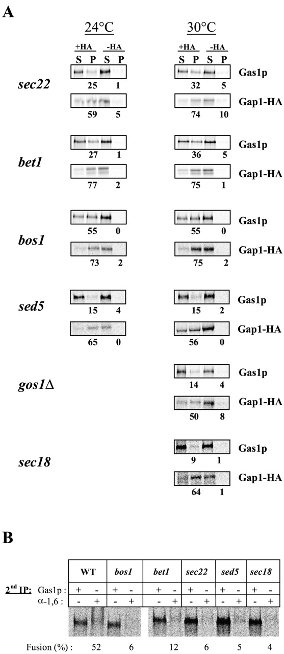
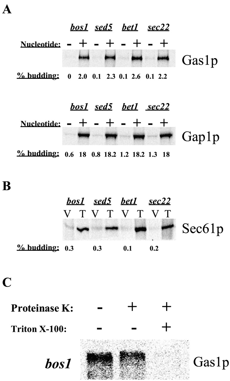
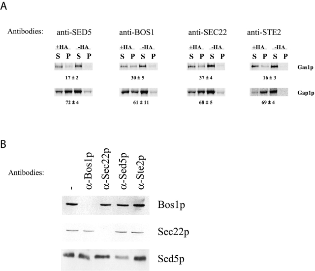
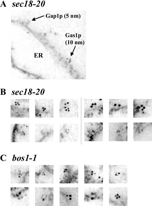
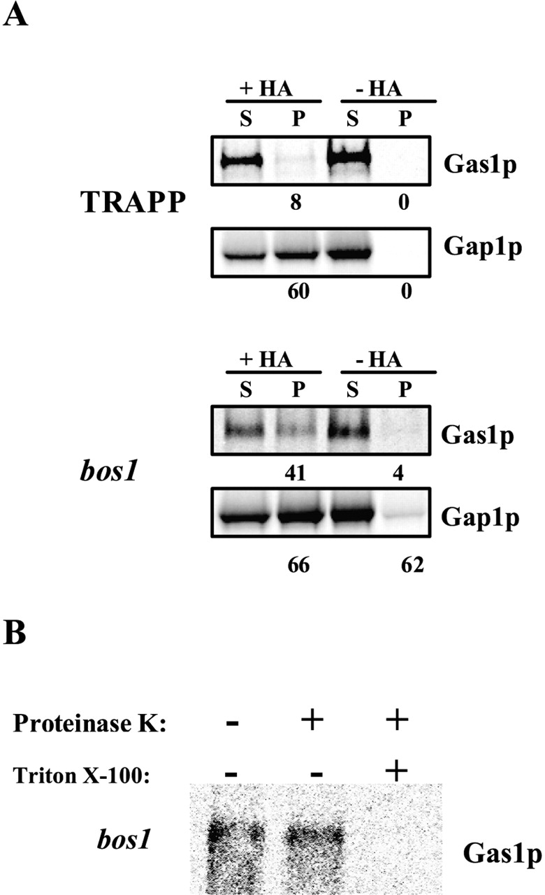
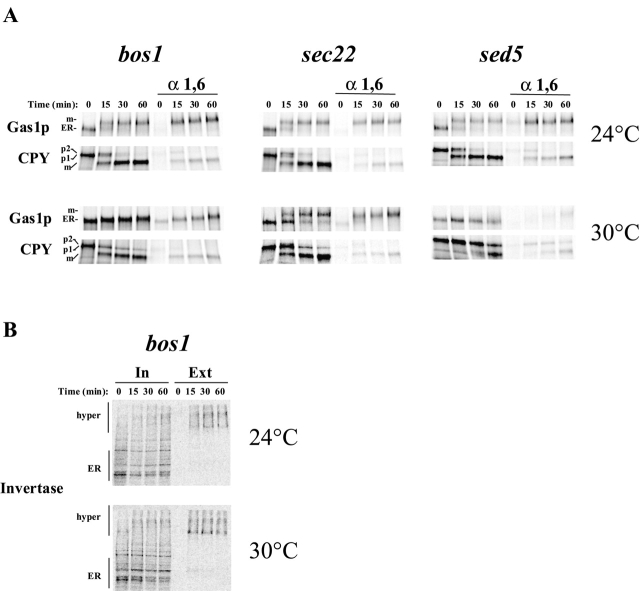
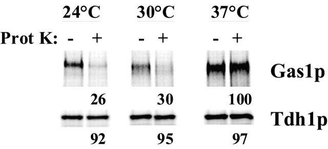
Glycosylphosphatidylinositol (GPI)-anchored proteins exit the ER in distinct vesicles from other secretory proteins, and this sorting event requires the Rab GTPase Ypt1p, tethering factors Uso1p, and the conserved oligomeric Golgi complex. Here we show that proper sorting depended on the vSNAREs, Bos1p, Bet1p, and Sec22p. However, the t-SNARE Sed5p was not required for protein sorting upon ER exit. Moreover, the sorting defect observed in vitro with bos1-1 extracts was also observed in vivo and was visualized by EM. Finally, transport and maturation of the GPI-anchored protein Gas1p was specifically affected in a bos1-1 mutant at semirestrictive temperature. Therefore, we propose that v-SNAREs are part of the cargo protein sorting machinery upon exit from the ER and that a correct sorting process is necessary for proper maturation of GPI-anchored proteins.


Figures

Figure 1.
Figure 1.
ER v-SNAREs are necessary for protein sorting upon ER exit. (A) Vesicles were generated from sec18–20 membranes and cytosol or from bos1–1, bet1–1, sec22–3, sed5–1, or gos1Δ membranes and wild-type cytosol. The vesicles were immunoisolated with or without monoclonal anti-HA antibody. The supernatants (S) and pellets (P) were processed for immunoprecipitation, and samples were analyzed by SDS-PAGE and quantified using a phosphorimager. The total recovery, S plus P, was set to 100%. Numbers represent the percentage of recovery in the pellet. This experiment is representative of at least two independent experiments. (B) ER budding and fusion with the Golgi compartment from wild-type, bos1–1, bet1–1, sec22–3, sed5–1, and sec18–20 membranes. Purified vesicles were processed for two consecutive immunoprecipitations. Samples were immunoprecipitated first with antibodies against Gas1p, then reprecipitated with antibodies against Gas1p or α-1,6 mannose, analyzed by SDS-PAGE, and quantified using a phosphor- imager. The percentage of fusion is the percentage of Gas1p after two immunoprecipitations that was recovered after precipitation with antibodies against α-1,6 mannose.
Figure 2.
Figure 2.
In vitro budding of ER-derived vesicles containing secretory proteins from SNARE mutants. (A) In vitro budding reactions were performed using sec18–20 membranes and cytosol or from bos1–1, bet1–1, sec22–3, sed5–1, or gos1Δ membranes and wild-type cytosol. The nucleotide requirement was tested in the absence of exogenous ATP and GTP and in presence of apyrase (1.5 U/reaction). Budding efficiencies were calculated as the percentage of the total input that was recovered in the purified vesicles for each individual protein. (B) Budding efficiencies determined for Sec61p packaging into ER-derived vesicles generated from bos1–1, bet1–1, sec22–3, and sed5–1 extracts. Numbers represent the percentage of the total input (T) that was recovered in the purified vesicles (V). (C) Integrity of vesicles generated from bos1–1 membranes. Purified vesicles produced from bos1–1 membranes were incubated on ice for 30 min with or without 0.5 mg/ml proteinase K in the presence or absence of 1% Triton X-100. Samples were processed for Gas1p immunoprecipitation, analyzed by SDS-PAGE, and visualized using a phosphorimager.
Figure 3.
Figure 3.
Anti–v-SNARE antibodies affect sorting. (A) Vesicles were generated from wild-type membranes and cytosol in the absence or presence of 5 μl of anti-Bos1p, anti-Sed5p, anti-Sec22p, or anti-Ste2p antibodies. The vesicles were immunoisolated with or without monoclonal anti-HA antibody. The supernatants (S) and pellets (P) were processed for immunoprecipitation, and samples were analyzed as described above. The total recovery, S plus P, was set to 100%. Numbers represent the percentage of recovery in the pellet from which the background was subtracted. The data are the mean ± SD of three independent experiments for anti-Bos1p, anti-Sed5p, and anti-Sec22p antibodies and two independent experiments for anti-Ste2p antibodies. (B) Immunoblot of floated vesicles generated from wild-type membranes and cytosol in absence or in presence of 5 μl of anti-Bos1p, anti-Sed5p, anti-Sec22p, or anti-Ste2p antibodies.
Figure 4.
Figure 4.
Immuno-EM of Gas1p and Gap1p upon exit from the ER in sec18–20 and bos1–1 mutants. In vitro budding reactions were performed using sec18–20 membranes and cytosol (A and B) or from bos1–1 membranes and wild-type cytosol (C). Extracts were fixed 10 min after addition of cytosol and incubation at 30°C. Immunodetection and quantification were performed as described in Materials and methods. For the quantification, >60 sections were counted for each mutant. In A, Gap1p and Gas1p were labeled with 5- and 10-nm gold particles, respectively. In B, Gas1p was labeled with either 10-nm (three left) or 15-nm (three right) gold particles (top); Gap1p was labeled with either 5-nm (three left) or 10-nm (three right) gold particles (bottom). In C, Gap1p and Gas1p were labeled with 5- and 10-nm gold particles, respectively (top) or with 10- and 15-nm gold particles, respectively (bottom).
Figure 5.
Figure 5.
Bos1p is necessary for sorting in vivo. (A) After pulse–chase labeling, vesicles were purified through a Nycodenz® step gradient directly from Bet3p-depleted cells or bos1–1 mutant cells and then immunoisolated with or without monoclonal anti-HA antibody. The supernatants (S) and pellet (P) were processed for immunoprecipitation, and samples were analyzed as described above. The total recovery, S plus P, was set to 100%. Numbers represent the percentage recovery in the pellet. (B) The integrity of bos1–1-generated vesicles was tested as described above.
Figure 6.
Figure 6.
In vivo Gas1p maturation is specifically affected in bos1–1 mutant at 30°C. Protein maturation in bos1–1, sec22–3, and sed5–1 mutants at 24 and 30°C. (A) Wild-type and mutant cells were pulse labeled and chased at the indicated temperatures for the indicated times. Gas1p and CPY were immunoprecipitated from cell lysates and separated by SDS-PAGE and quantified using a phosphorimager. (B) Analysis of invertase secretion in bos1–1 mutant. Cells were pulse–chase labeled, and then the cells and medium were separated and analyzed separately for invertase by immunoprecipitation, SDS-PAGE, and visualization using a phosphorimager.
Figure 7.
Figure 7.
Gas1p reaches the cell surface in the bos1–1 mutant. Mutant bos1–1 cells were pulse labeled for 3 min and chased for 60 min. Cell walls were mildly digested and incubated with or without proteinase K as described. Proteinase K was inactivated and Gas1p and Tdh1p were immunoprecipitated from cell lysates and analyzed by SDS-PAGE and quantified using a phosphorimager.

Similar articles

Cited by

References

    1. Allan, B.B., B.D. Moyer, and W.E. Balch. 2000. Rab1 recruitment of p115 into a cis-SNARE complex: programming budding COPII vesicles for fusion. Science. 289:444–448. - PubMed
    1. Bagnat, M., S. Keranen, A. Shevchenko, and K. Simons. 2000. Lipid rafts function in biosynthetic delivery of proteins to the cell surface in yeast. Proc. Natl. Acad. Sci. USA. 97:3254–3259. - PMC - PubMed
    1. Cao, X., N. Ballew, and C. Barlowe. 1998. Initial docking of ER-derived vesicles requires Uso1p and Ypt1p but is independent of SNARE proteins. EMBO J. 17:2156–2165. - PMC - PubMed
    1. Carroll, K.S., J. Hanna, I. Simon, J. Krise, P. Barbero, and S.R. Pfeffer. 2001. Role of rab9 GTPase in facilitating receptor recruitment by tip47. Science. 292:1373–1376. - PubMed
    1. Chamberlain, L.H., R.D. Burgoyne, and G.W. Gould. 2001. SNARE proteins are highly enriched in lipid rafts in PC12 cells: implications for the spatial control of exocytosis. Proc. Natl. Acad. Sci. USA. 98:5619–5624. - PMC - PubMed

Publication types

MeSH terms