. 2003 Aug 4;162(3):391-401.
doi: 10.1083/jcb.200303085. Epub 2003 Jul 28.

Importin beta contains a COOH-terminal nucleoporin binding region important for nuclear transport

Affiliations

Importin beta contains a COOH-terminal nucleoporin binding region important for nuclear transport

Janna Bednenko et al. J Cell Biol. .

Abstract

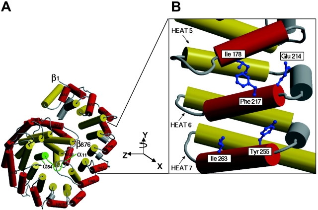
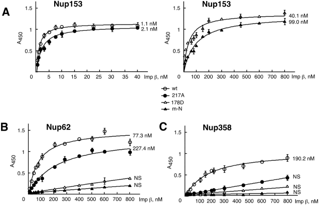
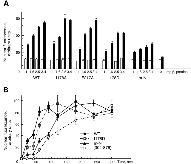
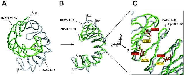
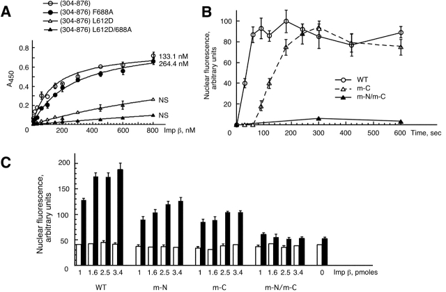
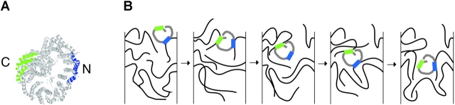
Proteins containing a classical NLS are transported into the nucleus by the import receptor importin beta, which binds to cargoes via the adaptor importin alpha. The import complex is translocated through the nuclear pore complex by interactions of importin beta with a series of nucleoporins. Previous studies have defined a nucleoporin binding region in the NH2-terminal half of importin beta. Here we report the identification of a second nucleoporin binding region in its COOH-terminal half. Although the affinity of the COOH-terminal region for nucleoporins is dramatically weaker than that of the NH2-terminal region, sets of mutations that perturb the nucleoporin binding of either region reduce the nuclear import activity of importin beta to a similar extent ( approximately 50%). An importin beta mutant with a combination of mutations in the NH2- and COOH-terminal regions is completely inactive for nuclear import. Thus, importin beta possesses two nucleoporin binding sites, both of which are important for its nuclear import function.


Figures

Figure 1.
Figure 1.
Model of the importin β–IBB domain complex. (A) Rod representation of the structure of importin β complexed with the IBB domain of importin α, according to Cingolani et al. (1999). The A and B helices of the HEAT repeats are shown in red and yellow, respectively, with connecting loops and helices in gray. The IBB domain of importin α is in green. (B) An enlarged view highlighting the A helices of HEAT repeats 5–7, which were shown to be involved in binding FxFG motifs by crystallography (Bayliss et al., 2000). The enlarged image was rotated by 90° in a Y direction with respect to A. The side chains of the residues analyzed by the mutagenesis as described in Table I are highlighted in blue.
Figure 2.
Figure 2.
Characterization of the binding of importin β to nucleoporins. Shown are binding isotherms depicting the interaction of selected importin β mutants with Nup153 (895–1475) (A), with full-length Nup62 (B), or with Nup358 (996–1963) (C). Error bars represent the standard deviation of duplicate measurements. The calculated apparent Kd values are indicated next to the binding isotherms. NS, nonsaturated binding at importin β concentrations up to 800 nM.
Figure 3.
Figure 3.
Effect of mutations in HEAT repeats 5–7 on nuclear import activity of importin β. (A) Nuclear import assays with wild-type importin β and with importin β mutants. 40-μl samples were incubated for 30 min at 30°C, and the level of nuclear import of FITC-labeled BSA-NLS was measured by flow cytometry. White bars depict control reactions performed in the presence of hexokinase and glucose to deplete ATP. Black bars show the levels of fluorescence in the presence of ATP. Error bars represent the standard deviation of duplicate measurements. (B) Kinetics of nuclear accumulation of importin β. 3.4 pmol of importin β or importin β (304–876) fragment was used in each 20-μl assay. The level of intranuclear importin β was quantified by immunofluorescence and confocal microscopy in samples fixed at the various time points. Error bars represent the standard deviation of two independent experiments. The level of intranuclear wild-type importin β for the 3-min time point was set at 100%, and all other values were normalized to the 100% level.
Figure 4.
Figure 4.
Structural alignment of the NH 2 - and COOH-terminal segments of importin β. (A) Worm representation of importin β. Residues 1–445 are highlighted in gray and residues 446–876 are in green. (B) Alignment of the NH2- and COOH-terminal segments of importin β. Residues 1–445 (HEAT repeats 1–10) are in gray and residues 446–876 (HEAT repeats 11–19) are in green. (C) The side chains of importin β residues I178, Y255, and I263 (yellow) closely align with those of L612, F688, and L695 (red), respectively. The enlarged image in C was rotated 90° in a Y direction and 30° in a Z direction with respect to B.
Figure 5.
Figure 5.
Effects of mutations in the COOH-terminal segment of importin β on nucleoporin binding and nuclear import. (A) Binding isotherms showing the interaction of the importin β fragment comprising residues 304–876 (with or without the mutations L612D and/or F688A) to Nup153 (895–1475). Error bars represent the standard deviation of duplicate measurements. The numbers next to the binding curves indicate apparent dissociation constants. NS, nonsaturated binding at importin β fragment concentrations of up to 800 nM. (B) Kinetics of nuclear accumulation of wild-type (WT), m-C, and m-N/m-C full-length importin β proteins. 3.4 pmol of each importin β construct was used in each 20-μl assay. The nuclear fluorescence was measured by immunofluorescence and confocal microscopy after 20–600 s incubation at room temperature, as indicated. Error bars represent the standard deviation of two independent experiments. The level of intranuclear wild-type importin β for the 3-min time point was set at 100%, and all other values were normalized as a fraction of the 100% level. (C) Nuclear import assays with the wild-type (WT), m-N, m-C, and m-N/m-C full-length importin β proteins. 40-μl reactions were incubated for 30 min at 30°C, and the level of nuclear import was measured by flow cytometry. White bars designate control reactions performed in the presence of hexokinase and glucose to deplete ATP. Black bars show the level of fluorescence in the presence of ATP. Error bars represent the standard deviation of duplicate measurements.
Figure 6.
Figure 6.
Model depicting the involvement of two nucleoporin binding regions in importin β in its translocation through the NPC. (A) The two nucleoporin binding regions are illustrated in a ribbon representation of importin β. The NH2-terminal region is shown in blue, and the COOH-terminal region in green. (B) One possible model for importin β translocation through the NPC. The model depicts only the central channel region, but movement between the peripheral fibrils of the NPC and the central channel could be conceptually similar. The two nucleoporin binding regions are shown in blue and green as in A, and the FG-rich nucleoporin regions are depicted as flexible filaments. Importin β could use its NH2- and COOH-terminal regions simultaneously or in succession to promote movement through the NPC.

References

    1. Adam, S.A., R. Sterne-Marr, and L. Gerace. 1992. Nuclear protein import using digitonin-permeabilized cells. Methods Enzymol. 219:97–110. - PubMed
    1. Bayliss, R., K. Ribbeck, D. Akin, H.M. Kent, C.M. Feldherr, D. Görlich, and M. Stewart. 1999. Interaction between NTF2 and xFxFG-containing nucleoporins is required to mediate nuclear import of RanGDP. J. Mol. Biol. 293:579–593. - PubMed
    1. Bayliss, R., T. Littlewood, and M. Stewart. 2000. Structural basis for the interaction between FxFG nucleoporin repeats and importin β in nuclear trafficking. Cell. 102:99–108. - PubMed
    1. Bayliss, R., T. Littlewood, L.A. Strawn, S.R. Wente, and M. Stewart. 2002. GLFG and FxFG nucleoporins bind to overlapping sites on importin β. J. Biol. Chem. 277:50597–50606. - PubMed
    1. Bednenko, J., G. Cingolani, and L. Gerace. 2003. Nucleocytoplasmic transport: navigating the channel. Traffic. 4:127–135. - PubMed

Publication types

MeSH terms