. 2003 Aug 4;162(3):443-55.
doi: 10.1083/jcb.200304008. Epub 2003 Jul 28.

Infectious HIV-1 assembles in late endosomes in primary macrophages

Affiliations

Infectious HIV-1 assembles in late endosomes in primary macrophages

Annegret Pelchen-Matthews et al. J Cell Biol. .

Abstract

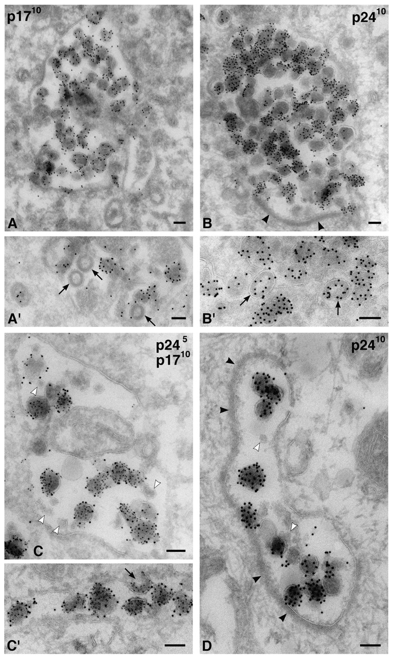
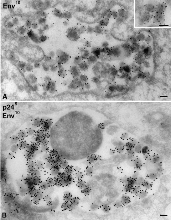
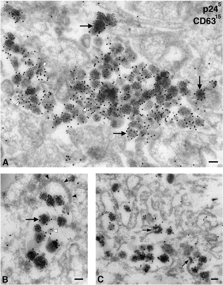
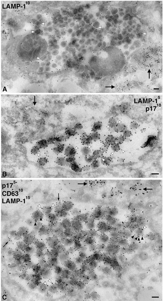
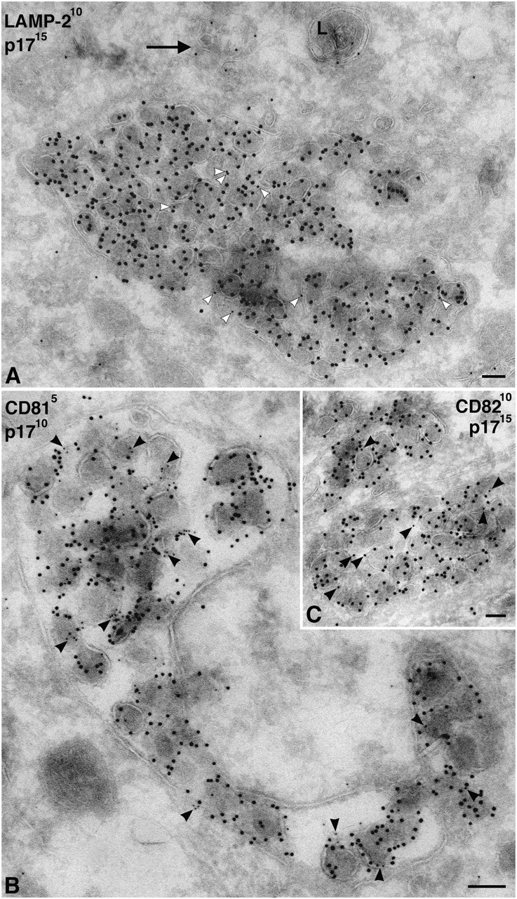
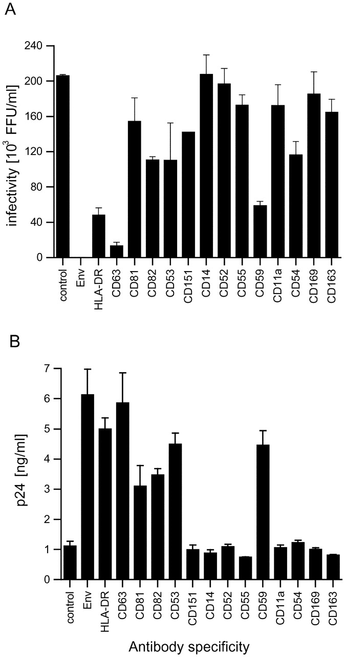
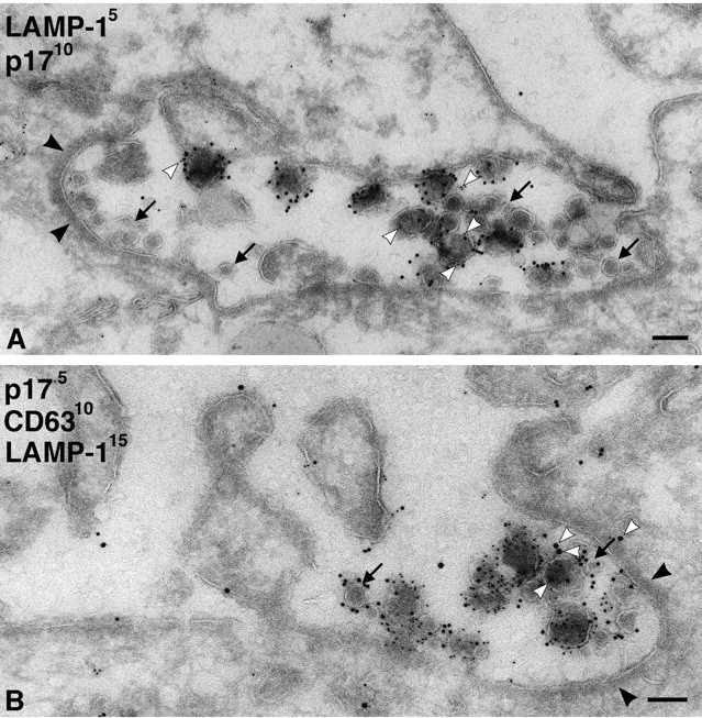
Although human immunodeficiency virus type 1 (HIV-1) is generally thought to assemble at the plasma membrane of infected cells, virions have been observed in intracellular compartments in macrophages. Here, we investigated virus assembly in HIV-1-infected primary human monocyte-derived macrophages (MDM). Electron microscopy of cryosections showed virus particles, identified by their morphology and positive labeling with antibodies to the viral p17, p24, and envelope proteins, in intracellular vacuoles. Immunolabeling demonstrated that these compartments contained the late endosomal marker CD63, which was enriched on vesicles within these structures and incorporated into the envelope of budding virions. The virus-containing vacuoles were also labeled with antibodies against LAMP-1, CD81, and CD82, which were also incorporated into the viral envelope. To assess the cellular source of infectious viruses derived from MDM, virus-containing media from infected cells were precipitated with specific antibodies. Only antibodies against antigens found in late endosomes precipitated infectious virus, whereas antibodies against proteins located primarily on the cell surface did not. Our data indicate that most of the infectious HIV produced by primary macrophages is assembled on late endocytic membranes and acquires antigens characteristic of this compartment. This notion has significant implications for understanding the biology of HIV and its cell-cell transmission.


Figures

Figure 1.
Figure 1.
Time course of HIV-1 Ba-L production in MDM. Human MDM were infected with 9.3 × 10⁵ focus-forming units (FFU) of HIV-1 Ba-L. Supernatants were collected daily and analyzed for (A) p24 content (○) or reverse transcriptase (•) and (B) infectivity on NP-2 CD4/CCR5 indicator cells. Activities are corrected for the dilution effect of feeding.
Figure 2.
Figure 2.
Overview of anti-p24 and -p17 staining on HIV-infected MDM. Semi-thin cryosections of MDM infected with HIV-1 Ba-L and labeled with anti-p24 (A–D), or infected with HIV-1 SF162 and labeled with anti-p17 (E–G), were stained with Alexa® Fluor 488 goat anti–mouse IgG. Cells had been infected for 7 (A), 12 (B), 15 (C), and 20 d (D); or 14 (E), 21 (F), and 30 d (G). Bars, 10 μm.
Figure 3.
Figure 3.
Intracellular HIV-1 in MDM. Cryosections from MDM infected with HIV-1 Ba-L were stained with anti-p17 (A and A′) or anti-p24 antibodies (B and B′) and 10 nm PAG. Alternatively, sections were double labeled for p24 (PAG 5 nm) and p17 (PAG 10 nm) (C and C′). Virus particles were found primarily in intracellular vacuoles at all times, i.e., in these cells infected for 7 (B′), 12 (A′), 14 (A and B), or 20 d (C and C′). Arrows identify budding or immature virions in A′, B′, or C′. D shows a cell infected with HIV-1 SF162 for 14 d, stained with anti-p24 and PAG10. Note the small unlabeled internal vesicles (C and D, white arrowheads) and the flat coat on some of these vacuoles (B and D, black arrowheads). Bars, 100 nm.
Figure 4.
Figure 4.
Detection of HIV-1 envelope on intracellular HIV-1 virions in MDM. Cryosections from MDM infected with HIV-1 Ba-L for 14 d were stained with anti-Env 2G12 and PAG10 (A) or double labeled for p24 with PAG5 and Env with PAG10 (B). The inset in A shows a detailed view with gold particles over the viral membrane on equatorially sectioned virions, or all over virions cut more tangentially. Bars, 100 nm.
Figure 5.
Figure 5.
HIV-1 virions in MDM accumulate in CD63-containing compartments. Cryosections from MDM infected with HIV-1 Ba-L for 14 d were double labeled for p24 with PAG5 and for CD63 with PAG15. (A) Overview of a large complex intracellular vacuole filled with virions and clusters of small vesicles containing CD63 (white arrowheads). Virions are identified by PAG5 and are frequently labeled with the larger gold particles marking CD63 (e.g., at the large arrows), whereas the small vesicles are labeled only with CD63/PAG15. (B) A smaller vacuole containing virus particles also labeled with CD63/PAG15. Note the electron-dense coat (black arrowheads). (C) Virions budding into a complex vacuole labeled with anti-CD63. Small arrows indicate budding virus particles incorporating CD63 into their membranes. Bars, 100 nm.
Figure 6.
Figure 6.
Virus-containing vacuoles in MDM labeled with antibodies to LAMP-1. (A) Cryosections from MDM infected with HIV-1 Ba-L for 7 d were labeled with a rabbit antiserum to LAMP-1 and PAG10. Note gold labeling associated with the large virus-containing vacuole, including gold particles on individual virions (white arrowheads). However, lysosomal membranes nearby contain significantly more gold particles (arrows). (B) Cryosection double labeled with rabbit anti-LAMP-1/PAG5 and anti-p17/PAG10. The large virus- containing vacuole contains many p17-labeled virions, whereas a few 5 nm gold particles identify associated LAMP-1 (e.g., at the white arrowheads). More strongly LAMP-1–labeled membranes are observed nearby (arrow). (C) A large virus vacuole triple labeled with anti-p17/PAG5, anti-CD63/PAG10, and rabbit anti-LAMP-1/PAG15. Many virions are labeled with the anti-CD63/PAG10 (black arrowheads), and some also contain a few LAMP-1/PAG15 particles, some of which are identified by white arrowheads. The rare, small internal vesicles contain just CD63 (small arrows). CD63 and LAMP-1–labeled membranes are also observed nearby (large arrows). Bars, 100 nm.
Figure 7.
Figure 7.
Cellular markers associated with virus-containing vacuoles in MDM. (A) Cryosections of MDM infected with HIV-1 Ba-L were double labeled with rabbit anti-LAMP-2 and PAG10, and virions were identified with anti-p17/PAG15. LAMP-2 was not highly expressed on these MDM, and hence only few gold particles could be observed, but there was some labeling of virus-containing vacuoles (white arrowheads). In addition, a lysosome (L) and membrane tubules are labeled nearby (arrow). (B and C) Cryosections of MDM infected with HIV-1 Ba-L were double labeled with antibodies against CD81 (B) or CD82 (C), whereas virions were identified with anti-p17. Both CD81 and CD82 are associated with the virus-containing vacuoles and can sometimes be found on the virions (black arrowheads). Labeling with anti-CD81 is significantly stronger than with anti-CD82. Bars, 100 nm.
Figure 8.
Figure 8.
Virus precipitation with antibodies against cellular proteins. Cell-free supernatants of MDM-derived HIV-1 Ba-L were precipitated with the antibodies indicated. Unprecipitated supernatants were analyzed for remaining infectious virus by a focus-forming assay on NP-2 CD4/CCR5 cells (A). Pellets were analyzed for precipitated viral protein by p24 ELISA (B).
Figure 9.
Figure 9.
Exocytosis of virions on infected MDM. Plasma membrane invaginations probably resulting from the fusion of intracellular virus-containing vacuoles with the plasma membrane. Cryosection in A was double labeled for LAMP-1/PAG5 and p17/PAG10, whereas B was triple labeled for p17/PAG5, CD63/PAG10, and LAMP-1/PAG15. Although these invaginations are continuous with the cell surface, they show labeling for LAMP-1 (white arrowheads) and CD63. Virions are interspersed with many small vesicles (black arrows) that are strongly labeled for CD63 (B, black arrows). Also, note the thick electron-dense coats on some parts of these invaginations (black arrowheads). Bars, 100 nm.

References

    1. Andrews, N.W. 2000. Regulated secretion of conventional lysosomes. Trends Cell Biol. 10:316–321. - PubMed
    1. Barre-Sinoussi, F., J.C. Chermann, F. Rey, M.T. Nugeyre, S. Chamaret, J. Gruest, C. Dauguet, C. Axler-Blin, F. Vezinet-Brun, C. Rouzioux, et al. 1983. Isolation of a T-lymphotropic retrovirus from a patient at risk for acquired immune deficiency syndrome (AIDS). Science. 220:868–871. - PubMed
    1. Blott, E.J., and G.M. Griffiths. 2002. Secretory lysosomes. Nat. Rev. Mol. Cell Biol. 3:122–131. - PubMed
    1. Boes, M., J. Cerny, R. Massol, M. Op den Brouw, T. Kirchhausen, J. Chen, and H.L. Ploegh. 2002. T-cell engagement of dendritic cells rapidly rearranges MHC class II transport. Nature. 418:983–988. - PubMed
    1. Bourinbaiar, A.S., and D.M. Phillips. 1991. Transmission of human immunodeficiency virus from monocytes to epithelia. J. Acquir. Immune Defic. Syndr. 4:56–63. - PubMed

Publication types

MeSH terms