. 2003 Aug;77(16):8661-8.
doi: 10.1128/jvi.77.16.8661-8668.2003.

Nonstructural proteins NS1 and NS2 of bovine respiratory syncytial virus block activation of interferon regulatory factor 3

Affiliations

Nonstructural proteins NS1 and NS2 of bovine respiratory syncytial virus block activation of interferon regulatory factor 3

Birgit Bossert et al. J Virol. 2003 Aug.

Abstract

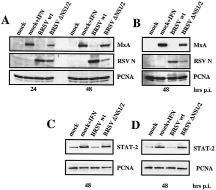
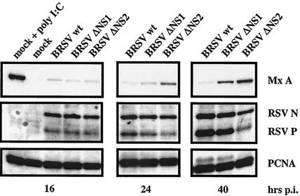
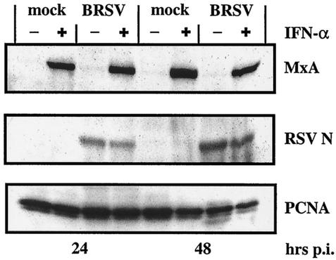
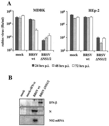
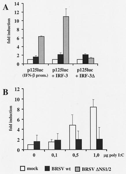
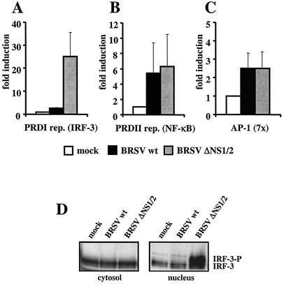
We have previously shown that the nonstructural (NS) proteins NS1 and NS2 of bovine respiratory syncytial virus (BRSV) mediate resistance to the alpha/beta interferon (IFN)-mediated antiviral response. Here, we show that they, in addition, are able to prevent the induction of beta IFN (IFN-beta) after virus infection or double-stranded RNA stimulation. In BRSV-infected MDBK cells upregulation of IFN-stimulated genes (ISGs) such as MxA did not occur, although IFN signaling via JAK/STAT was found intact. In contrast, infection with recombinant BRSVs lacking either or both NS genes resulted in efficient upregulation of ISGs. Biological IFN activity and IFN-beta were detected only in supernatants of cells infected with the NS deletion mutants but not with wild-type (wt) BRSV. Subsequent analyses of IFN-beta promoter activity showed that infection of cells with the double deletion mutant BRSV DeltaNS1/2, but not with BRSV wt, resulted in a significant increase in IFN-beta gene promoter activity. Induction of the IFN-beta promoter depends on the activation of three distinct transcription factors, NF-kappaB, ATF-2/c-Jun, and IFN regulatory factor 3 (IRF-3). Whereas NF-kappaB and ATF-2/c-Jun activities were readily detectable and comparable in both wt BRSV- and BRSV DeltaNS1/2-infected cells, phosphorylation and transcriptional activity of IRF-3, however, were observed only after BRSV DeltaNS1/2 infection. NS protein-mediated inhibition of IRF-3 activation and IFN induction should have considerable impact on the pathogenesis and immunogenicity of BRSV.


Figures

FIG. 1.
FIG. 1.
BRSV NS deletion mutants but not wt BRSV induce ISGs. Shown are Western blot analyses of MxA expression in MDBK (A) and HEp-2 (B) cells and of STAT-2 in MDBK (C) and HEp-2 (D) cells. Cells were mock infected or infected at an MOI of 0.2 with BRSV wt and BRSV ΔNS1/2. As a control for upregulation of ISGs, mock-infected cells were treated with 2,000 U of recombinant IFN-α A/D per ml. Cells were harvested 24 and 48 h postinfection as indicated, and equal amounts of cell extract were subjected to SDS-PAGE followed by detection of MxA, STAT-2, BRSV N protein, and PCNA as a loading control.
FIG. 2.
FIG. 2.
Induction of MxA is delayed in BRSV ΔNS1-infected cells. MDBK cells were mock infected or infected at an MOI of 0.2 with BRSV wt, BRSV ΔΝS1, or BRSV ΔΝS2. When indicated, 2,000 U of IFN-α A/D per ml was added immediately after infection. Cells were harvested 16, 24, and 40 h postinfection, and equal amounts of cell extract were subjected to SDS-PAGE followed by detection of MxA, BRSV N and P protein, and PCNA.
FIG. 3.
FIG. 3.
BRSV wt does not interfere with IFN signaling and induction of ISGs by IFN. MDBK cells were mock infected or infected at an MOI of 0.2 with BRSV wt. When indicated, 2,000 U of IFN-α A/D per ml was added immediately after infection. Cells were harvested 24 or 48 h postinfection, and equal amounts of cell extract were subjected to SDS-PAGE followed by detection of MxA, BRSV N protein, and PCNA.
FIG. 4.
FIG. 4.
(A) IFN activity in supernatants of infected MDBK and HEp-2 cells. Cells were mock infected or infected at an MOI of 0.2 with BRSV wt or BRSV ΔNS1/2. Supernatants were harvested 24, 48, and 72 h postinfection and transferred onto RV-infected MDBK cells. RV titers were determined 2 days postinfection. Error bars indicate standard deviations. (B) IFN-β is expressed in BRSV ΔNS1/2-infected but not in wt BRSV-infected HEp-2 cells. Supernatant proteins were precipitated with TCA, and IFN-β was identified in a Western blot made with a polyclonal anti-human IFN-β serum. RSV N protein in cell extracts and NS2 mRNA in total RNA were demonstrated by Western blotting with an anti-RSV serum and an NS2-specific cDNA hybridization probe, respectively, at 37 h postinfection.
FIG. 5.
FIG. 5.
Virus-induced activation of the IFN-β enhancer. (A) HEK 293 cells were transfected with a luciferase construct driven by the IFN-β promoter-enhancer (p125luc) and, as indicated, cotransfected with plasmids encoding IRF-3 or a dominant-negative IRF-3 mutant (ΔIRF-3). Ten hours posttransfection cells were infected at an MOI of 0.2 with BRSV wt or BRSV ΔNS1/2. Cell lysates were harvested 14 h postinfection and assayed for luciferase activity. Relative light units are given as fold induction and represent the mean values of four independent experiments with error bars indicating standard deviations. (B) BRSV wt inhibits IFN induction triggered by poly(rI):poly(rC). HEK 293 cells were mock infected or infected with BRSV wt at an MOI of 0.25. Twelve hours postinfection, cells were transfected with p125luc and the indicated amounts of poly(rI):poly(rC). Luciferase assays were done 24 h posttransfection. Relative light units are given as fold induction and represent the mean values of three independent experiments. Error bars indicate standard deviations.
FIG. 6.
FIG. 6.
BRSV wt selectively blocks the activation of IRF-3. (A to C) HEK 293 cells were transfected with plasmids harboring the luciferase gene under the control of promoters containing either IRF-3 (A), NF-κB (B), or AP-1 (C) binding sites. Ten hours posttransfection, cells were infected with the indicated viruses at an MOI of 0.2. Fourteen hours postinfection cells were harvested and luciferase activity was determined. Relative light units are given as fold induction. Results show the mean values of four independent experiments with error bars showing standard deviations. (D) IRF-3 is not phosphorylated in BRSV wt-infected cells. HEK 293 cells were mock infected or infected with the indicated viruses at an MOI of 0.25. Ten hours postinfection, cells were harvested, and nuclear extracts were prepared as described in Materials and Methods and subjected to SDS-PAGE. Nonphosphorylated and phosphorylated forms of IRF-3 were detected with an anti-IRF-3 antibody (Dianova).

Similar articles

Cited by

References

    1. Aebi, M., J. Fah, N. Hurt, C. E. Samuel, D. Thomis, L. Bazzigher, J. Pavlovic, O. Haller, and P. Staeheli. 1989. cDNA structures and regulation of two interferon-induced human Mx proteins. Mol. Cell. Biol. 9:5062-5072. - PMC - PubMed
    1. Akira, S., K. Takeda, and T. Kaisho. 2001. Toll-like receptors: critical proteins linking innate and acquired immunity. Nat. Immunol. 2:675-680. - PubMed
    1. Alexopoulou, L., A. C. Holt, R. Medzhitov, and R. A. Flavell. 2001. Recognition of double-stranded RNA and activation of NF-κB by Toll-like receptor 3. Nature 413:732-738. - PubMed
    1. Andrejeva, J., D. F. Young, S. Goodbourn, and R. E. Randall. 2002. Degradation of STAT1 and STAT2 by the V proteins of simian virus 5 and human parainfluenza virus type 2, respectively: consequences for virus replication in the presence of alpha/beta and gamma interferons. J. Virol. 76:2159-2167. - PMC - PubMed
    1. Atreya, P. L., M. E. Peeples, and P. L. Collins. 1998. The NS1 protein of human respiratory syncytial virus is a potent inhibitor of minigenome transcription and RNA replication. J. Virol. 72:1452-1461. - PMC - PubMed

Publication types

MeSH terms

LinkOut - more resources