. 2003 Aug;77(16):8686-94.
doi: 10.1128/jvi.77.16.8686-8694.2003.

PML residue lysine 160 is required for the degradation of PML induced by herpes simplex virus type 1 regulatory protein ICP0

Affiliations

PML residue lysine 160 is required for the degradation of PML induced by herpes simplex virus type 1 regulatory protein ICP0

Chris Boutell et al. J Virol. 2003 Aug.

Abstract

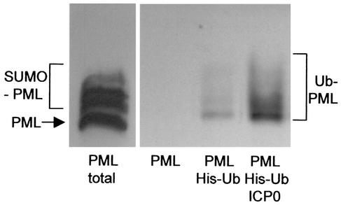
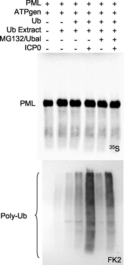
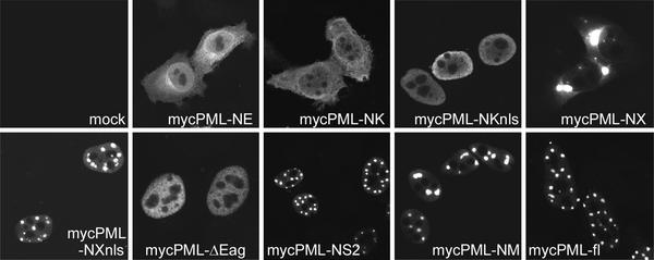
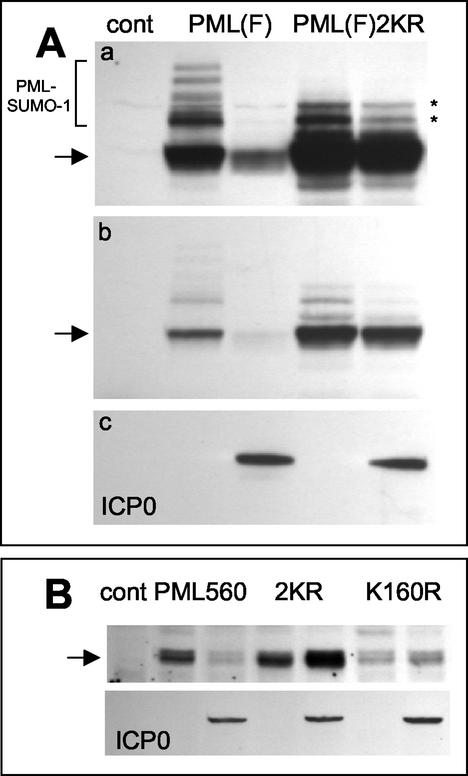
During the early stages of herpes simplex virus type 1 (HSV-1) infection, viral immediate-early regulatory protein ICP0 localizes to and disrupts cellular nuclear structures known as PML nuclear bodies or ND10. These activities correlate with the functions of ICP0 in stimulating lytic infection and reactivating quiescent HSV-1. The disruption of ND10 occurs because ICP0 induces the loss of the SUMO-1-modified forms of PML and the subsequent proteasome-mediated degradation of the PML protein. The functions of ICP0 are largely dependent on the integrity of its zinc-binding RING finger domain. Many RING finger proteins have been found to act as ubiquitin E3 ligase enzymes, stimulating the production of conjugated polyubiquitin chains in the presence of ubiquitin, the ubiquitin-activating enzyme E1, and the appropriate E2 ubiquitin-conjugating enzyme. Substrate proteins that become polyubiquitinated are then subject to degradation by proteasomes. We have previously shown that purified full-length ICP0 acts as an efficient E3 ligase in vitro, producing high-molecular-weight polyubiquitin chains in a RING finger-dependent but substrate-independent manner. In this paper we report on investigations into the factors governing the degradation of PML induced by ICP0 in a variety of in vivo and in vitro assays. We found that ICP0 expression increases the levels of ubiquitinated PML in transfected cells. However, ICP0 does not interact with or directly ubiquitinate either unmodified PML or SUMO-1-modified PML in vitro, suggesting either that additional factors are required for the ICP0-mediated ubiquitination of PML in vivo or that PML degradation is an indirect consequence of some other activity of ICP0 at ND10. Using a transfection-based approach and a family of deletion and point mutations of PML, we found that efficient ICP0-induced PML degradation requires sequences within the C-terminal part of PML and lysine residue 160, one of the principal targets for SUMO-1 modification of the protein.


Figures

FIG. 1.
FIG. 1.
ICP0 induces the formation of ubiquitinated PML in transfected cells. HEp-2 cells were transfected with combinations of plasmids expressing epitope-tagged PML, polyhistidine-tagged ubiquitin with the lysine 48-to-arginine (K48R) mutation (His-Ub), and ICP0 (as indicated below the panels). The left panel shows a sample of a whole-cell extract of cells transfected with the PML expression plasmid alone (with the unmodified and SUMO-1-modified forms of PML indicated). The right panel shows a Western blot of ubiquitinated total cell proteins that had been purified by metal chelate affinity chromatography as described in Materials and Methods and probed for PML. PML was not detected in the left lane of this panel, which is a control with proteins purified from cells transfected with the PML plasmid in the absence of the tagged K48R ubiquitin plasmid. Inclusion of this plasmid results in the isolation of ubiquitin-modified PML bands (center lane). The quantity of these bands was greatly increased in the presence of coexpressed ICP0 (right lane).
FIG. 2.
FIG. 2.
ICP0 does not ubiquitinate unmodified PML in vitro. (A) The ubiquitin-activating enzyme E1, ICP0, and various E2 ubiquitin-conjugating enzymes were expressed, purified, and mixed in a buffer containing ATP and ubiquitin (Ub). The products of these substrate-independent reactions were analyzed by Western blotting, with probing for the formation of high-molecular-weight polyubiquitin chains by using monoclonal antibody FK2. Such chains were readily produced in the presence of UbcH5a and UbcH6 but not with the other E2 enzymes tested. (B) PML protein was produced by coupled in vitro transcription-translation and added to reaction mixtures containing ICP0, UbcH5a, and the other basic components of the ubiquitin conjugation machinery. There was no evidence of the formation of ubiquitinated PML species with lower gel mobility.
FIG. 3.
FIG. 3.
ICP0 does not ubiquitinate unmodified PML in the presence of a mixture of endogenous cellular ubiquitination enzymes. PML was produced by coupled in vitro transcription-translation and mixed with ICP0, an ATP generating system (ATPgen), the proteasome inhibitor MG132 and ubiquitin aldehyde (to inhibit proteasomes and ubiquitin-specific proteases, respectively), and an extract containing a mixture of endogenous ubiquitin conjugating enzymes (Ub Extract). No ubiquitination of PML was observed (upper panel) despite ICP0 inducing increased amounts of conjugated ubiquitin (lower panels).
FIG. 4.
FIG. 4.
ICP0 does not hinder the conjugation of SUMO-1 to PML, affect the stability of SUMO-1-modified PML, or ubiquitinate SUMO-1-modified PML in vitro. (A) In vitro-expressed PML was mixed with purified components of the SUMO-1 modification pathway, i.e., Sae1/Sae2, Ubc9, and SUMO-1. SUMO-1-modified PML was readily observed, as indicated by the bracketed bands above the labeled major unmodified PML band. The modified and unmodified forms of PML are similarly marked in panels B and C. (B) SUMO-1-modified PML was produced in the absence (lane 3) or presence (lanes 4 to 6) of ICP0. The reaction mixtures in lanes 1 to 6 were incubated for 180 min. In lanes 7 to 10, similar reaction mixtures were incubated for 180 min in the absence of ICP0 and then ICP0 was added to the reaction mixtures in lanes 8 to 10 and the incubations were continued for a further 180 min. ICP0 affected neither the efficiency of formation nor the stability of SUMO-1-modified PML. (C) ICP0 does not ubiquitinate SUMO-1-modified PML in vitro. PML was expressed in vitro and then mixed with the components of the SUMO-1 modification pathway, ICP0, UbcH6, and the other components of the ubiquitination pathway, as indicated. There was no evidence for the formation of additional ubiquitinated PML species (left panel), despite ICP0 producing polyubiquitin chains in these reactions, as detected by probing of parallel reactions for conjugated ubiquitin by using monoclonal antibody FK2 (right panel). Mock TnT in the right panel refers to the use of an amount of the TnT wheat germ transcription-translation mixture equivalent to that in the lanes in the left panel in the absence of added substrate plasmid and labeled methionine.
FIG. 5.
FIG. 5.
Map of PML isoform IV (633 amino acid residues [aa]), showing the RING finger (R), the two B boxes (B1 and B2), the coiled-coil region (CC), the nuclear localization signal (nls), and the two major characterized sites of SUMO-1 modification, lysine 160 and lysine 490. At the top are shown the positions of restriction sites in the cDNA that were used for the generation of the truncated derivatives depicted below. The addition of nls to the name of a truncated plasmid indicates that the simian virus 40 large-T-antigen nuclear localization signal had been added to the C-terminal end of the open reading frame.
FIG. 6.
FIG. 6.
Typical immunofluorescent localizations in transfected cells expressing the PML proteins depicted in Fig. 5.
FIG. 7.
FIG. 7.
Degradation of SUMO-1-modified and unmodified forms of PML by cotransfection expression of ICP0. The extracts were prepared 20 h after transfection. (A) Top panel, Western blot detection of Myc-tagged PML and deletion mutant variants with monoclonal antibody 9E10. The left lane shows a control (cont) whole-cell extract from cells transfected with vector plasmids. The other lanes show pairs of extracts from cells transfected with the indicated PML plasmid (see Fig. 5 for their structures) either without or with an ICP0 expression plasmid (on the left and right of each pair, respectively). The arrows indicate the positions of the unmodified forms of full-length PML and the NM and NS2 deletions. The modified forms of these proteins are of lesser intensity and migrate more slowly. The SUMO-1-modified forms of full-length PML are marked by the bracket. The modified forms of the truncated proteins have correspondingly increased mobilities. Although the modified PML isoforms expressed by each construct are subject to ICP0-induced degradation, the unmodified forms of the deletion mutants are relatively stable compared to the full-length protein. Middle panel, a lower exposure of the same blot, showing that the unmodified forms of the truncation mutants PML-NM and PML-NS2 are more resistant to ICP0-induced degradation that the parent full-length protein. Bottom panel, reprobing of the blot filter for ICP0, demonstrating equivalent expression levels in the three experiments. (B) PML truncation mutant PML-NK is stable in the presence of ICP0, and its level of expression is increased. The left three lanes are equivalent to the left trio of panel A, showing the modified and unmodified forms of full-length PML in the presence and absence of ICP0 as a control. The right four lanes show PML-NK expression levels in the absence of ICP0 and with increasing amounts of the ICP0 expression plasmid in the transfection mixture (20, 50, and 100 ng, as indicated by the wedge).
FIG. 8.
FIG. 8.
PML lysine residue 160, which is subject to conjugation to SUMO-1, is required for efficient ICP0-induced PML degradation. (A) Panel a, the left three lanes are equivalent to those in Fig. 7A, top panel, showing the modified (bracket) and unmodified (arrow) forms of full-length PML in the presence and absence of ICP0 as a control (cont). The right two lanes show the levels of expression of a PML derivative with lysine-to-arginine mutations at residues 160 and 490, the major characterized SUMO-1 modification sites of PML, in the presence and absence of coexpressed ICP0. The asterisks indicate uncharacterized modified forms of the mutant PML protein that migrate more rapidly than most of the SUMO-1-modified forms of the normal protein. Panel b, a shorter exposure of the same blot, showing the relative insensitivity of the double lysine mutant to ICP0 expression. Panel c, a reprobe of the same blot to detect ICP0 in the relevant lanes. (B) A similar experiment using the 560-residue PML isoform VI. The upper panel shows the control lane and then paired lanes of PML isoform VI (PML560), the double lysine mutant PML560SUMO- (2KR), and the single K160R mutant detected by Western blotting in extracts of cells cotransfected or not with an ICP0 expression plasmid (as detected in the lower panel by reprobing of the same filter). The amount of PML isoform VI was decreased by ICP0, whereas the amounts of the double and single lysine mutants were increased. The basal level of the single K160R mutant was lower than those of the other two proteins in this experiment.

Similar articles

Cited by

References

    1. Ahn, J. H., and G. S. Hayward. 2000. Disruption of PML-associated nuclear bodies by IE1 correlates with efficient early stages of viral gene expression and DNA replication in human cytomegalovirus infection. Virology 274:39-55. - PubMed
    1. Bailey, D., and P. O'Hare. 2002. Herpes simplex virus 1 ICP0 co-localizes with a SUMO-specific protease. J. Gen. Virol. 83:2951-2964. - PubMed
    1. Boutell, C., S. Sadis, and R. D. Everett. 2002. Herpes simplex virus type 1 immediate-early protein ICP0 and its isolated RING finger domain act as ubiquitin E3 ligases in vitro. J. Virol. 76:841-850. - PMC - PubMed
    1. Chelbi-Alix, M. K., and H. de The. 1999. Herpes virus induced proteasome-dependent degradation of the nuclear bodies-associated PML and Sp100 proteins. Oncogene 18:935-941. - PubMed
    1. Duprez, E., A. J. Saurin, J. M. Desterro, V. Lallemand-Breitenbach, K. Howe, M. N. Boddy, E. Solomon, H. de The, R. T. Hay, and P. S. Freemont. 1999. SUMO-1 modification of the acute promyelocytic leukaemia protein PML: implications for nuclear localisation. J. Cell Sci. 112:381-393. - PubMed

Publication types

MeSH terms

LinkOut - more resources