. 2003 Aug;77(16):8719-28.
doi: 10.1128/jvi.77.16.8719-8728.2003.

Induction of protective immune responses against R5 human immunodeficiency virus type 1 (HIV-1) infection in hu-PBL-SCID mice by intrasplenic immunization with HIV-1-pulsed dendritic cells: possible involvement of a novel factor of human CD4(+) T-cell origin

Affiliations

Induction of protective immune responses against R5 human immunodeficiency virus type 1 (HIV-1) infection in hu-PBL-SCID mice by intrasplenic immunization with HIV-1-pulsed dendritic cells: possible involvement of a novel factor of human CD4(+) T-cell origin

Atsushi Yoshida et al. J Virol. 2003 Aug.

Abstract

The potential of a dendritic cell (DC)-based vaccine against human immunodeficiency virus type 1 (HIV-1) infection in humans was explored with SCID mice reconstituted with human peripheral blood mononuclear cells (PBMC). HIV-1-negative normal human PBMC were transplanted directly into the spleens of SCID mice (hu-PBL-SCID-spl mice) together with autologous mature DCs pulsed with either inactivated HIV-1 (strain R5 or X4) or ovalbumin (OVA), followed by a booster injection 5 days later with autologous DCs pulsed with the same respective antigens. Five days later, these mice were challenged intraperitoneally with R5 HIV-1(JR-CSF). Analysis of infection at 7 days postinfection showed that the DC-HIV-1-immunized hu-PBL-SCID-spl mice, irrespective of the HIV-1 isolate used for immunization, were protected against HIV-1 infection. In contrast, none of the DC-OVA-immunized mice were protected. Sera from the DC-HIV-1- but not the DC-OVA-immunized mice inhibited the in vitro infection of activated PBMC and macrophages with R5, but not X4, HIV-1. Upon restimulation with HIV-1 in vitro, the human CD4(+) T cells derived from the DC-HIV-1-immunized mice produced a similar R5 HIV-1 suppressor factor. Neutralizing antibodies against human RANTES, MIP-1alpha, MIP-1beta, alpha interferon (IFN-alpha), IFN-beta, IFN-gamma, interleukin-4 (IL-4), IL-10, IL-13, IL-16, MCP-1, MCP-3, tumor necrosis factor alpha (TNF-alpha), or TNF-beta failed to reverse the HIV-1-suppressive activity. These results show that inactivated HIV-1-pulsed autologous DCs can stimulate splenic resident human CD4(+) T cells in hu-PBL-SCID-spl mice to produce a yet-to-be-defined, novel soluble factor(s) with protective properties against R5 HIV-1 infection.


Figures

FIG. 1.
FIG. 1.
Induction of antigen-specific human immune responses in hu-PBL-SCID-spl mice by immunization with antigen-pulsed DC. (A) PBMC (3 × 10⁶ cells) alone (nonimmune) (•), with OVA (100 μg) (OVA immune) (⧫), or with DC (5 × 10⁵ cells) pulsed with OVA (100 μg) (DC-OVA immune) (▪) were engrafted into the spleens of SCID mice. Five days later, the PBMC-OVA-immunized mice received a booster injection with OVA and the DC-OVA-immunized mice received a booster injection with DC-OVA. Five days later, serum samples were collected and human anti-OVA antibodies were measured by ELISA. Results are expressed as the mean ± standard deviation from six independent experiments. *, P < 0.05. (B) Lymphocytes (2 × 10⁶ cells) recovered from hu-PBL-SCID-spl mice were cocultured with 2 × 10⁵ autologous APC (adherent PBMC) in the presence (open bars) or absence (solid bars) of 1 μg of OVA per ml at37°C for 2 days in 1 ml of RPMI medium containing 20 U of human IL-2 per ml. APC cultured alone served as controls. Results are expressed as the mean ± standard deviation from six independent experiments. *, P < 0.05. (C) Lymphocytes (2 × 10⁶ cells) recovered from hu-PBL-SCID-spl mice which were immunized with either DC-OVA or DC-AT-2-inactivated HIV-1JR-CSF were either not restimulated (solid bars) or restimulated as outlined for panel B in the presence of OVA (open bars) or AT-2-inactivated HIV-1 (containing 40 ng of p24) (shaded bars), respectively, for 2 days. Supernatant fluids from such cultures were harvested, and the levels of human IFN-γ were determined by ELISA. All results are expressed as the mean ± standard deviation from six independent experiments. *, P < 0.05.
FIG. 2.
FIG. 2.
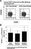
Human CD4+ T cells from HIV-1-protected hu-PBL-SCID-spl mice express CCR5 and are permissive for R5 HIV-1 infection in vitro. (A) Lymphocytes recovered from hu-PBL-SCID-spl mice immunized with DC-OVA or DC-HIV-1JR-CSF were stained with anti-CD4, anti-CCR5, and anti-CXCR4. The expression profiles of CCR5 and CXCR4 on the CD4+ T cells are shown. (B) Lymphocytes recovered from hu-PBL-SCID-spl mice immunized with DC-OVA, DC-HIV-1JR-CSF, or DC-HIV-1NL4-3 were washed and infected with 500 TCID50 of HIV-1JR-CSF at 37°C for 4 h in vitro. After washing, the cells were cultured for 5 days in RPMI medium containing 20 U of IL-2 per ml. Levels of HIV-1 p24 present in culture supernatants were quantitated by ELISA. All results are expressed as the mean ± standard deviation from six independent experiments.
FIG. 3.
FIG. 3.
Inhibition of R5 (A), but not X4 (B), HIV-1 infection by the HIV-1 immune serum. PBMC (5 × 10⁵ cells/well) activated in vitro for 3 days were washed and then incubated in medium (▪) or final 20% serum samples obtained from either DC-OVA-immune (⧫) or DC-HIV-1JR-CSF-immune (▴) hu-PBL-SCID-spl mice at 37°C for 1 h, followed by the addition of 500 TCID50 of HIV-1JR-CSF or HIV-1NL4-3 and further incubation at 37°C for 4 h. After washing, cells were incubated in IL-2-containing medium for 5 days. The level of HIV-1 replication was monitored by quantitating HIV-1 p24 levels in the culture supernatants. Results are expressed as the mean ± standard deviation from six independent experiments.
FIG. 4.
FIG. 4.
Inhibition of R5, but not X4, HIV-1 by the HIV-1 immune serum. In vitro-activated PBMC were treated at 37°C for 1 h with 10% pooled serum samples obtained from DC-HIV-1JR-CSF-immune hu-PBL-SCDI-spl mice, followed by the addition of 500 TCID50 of HIV-1 (R5 isolates JR-CSF, JR-FL, and SF162 and X4 isolates NL4-3 and IIIB) and further incubation at 37°C for 4 h. After washing, cells were incubated in IL-2-containing medium for 5 days, and levels of HIV-1 p24 in the culture supernatants were quantitated. The HIV-1-suppressive activities of the serum preincubated with the mixture of anti-β-chemokine antibodies (+Ab) were also determined. Percent inhibition was calculated by using values obtained with the medium controls, as follows: JR-CSF, 18.7 ng/ml; JR-FL, 7.6 ng/ml; SF162, 6.6 ng/ml; NL4-3, 18.3 ng/ml; and IIIB, 10.2 ng/ml.
FIG. 5.
FIG. 5.
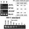
Blocking of R5 HIV-1 infection in human macrophage cultures. Cultured macrophages or activated PBMC were preincubated in medium alone (lane 1) or with pooled serum samples from DC-HIV-1JR-CSF-immune SCID mice in the absence (lane 2) or presence (lane 3) of a mixture of anti-β chemokines, and then the macrophages were infected with R5 HIV-1 strains (JR-FL, SF-162, and JR-CSF) and the PBMC were infected with X4 HIV-1NL4-3. After washing, the cells were cultured for 2 days and cellular DNA were extracted and analyzed for the number of HIV-1 provirus copies. The signal obtained for actin was utilized as a reference control. The estimated numbers of HIV-1 copies per 10,000 copies of actin are shown in the accompanying table. The data shown are representative of those from three independent experiments.
FIG. 6.
FIG. 6.
Identification of the cell population producing the suppressive factor. (A) Lymphocytes from DC-HIV-1JR-CSF-immune hu-PBL-SCID-spl mice were positively selected into human CD4+ and CD8+ T-cell subpopulations by using anti-CD4 and anti-CD8 MAb-conjugated immunobeads. Unfractionated (lymphocytes) or CD4+ or CD8+ T-cell populations (2 × 10⁶ cells) were cocultured with autologous APC (2 × 10⁵ cells) in the presence of AT-2-inactivated HIV-1 (containing 40 ng of p24) in 1 ml of IL-2-containing medium. After 2 days, culture supernatants were harvested to quantitate the levels of HIV-1 suppressor activity. Activated PBMC (target cells) were pretreated with these culture supernatants (50% final concentration) in the absence or presence (+Ab) of a mixture of anti-β-chemokine neutralizing antibodies and then infected with 500 TCID50 of either HIV-1JR-CSF (solid bars) or HIV-1NL4-3 (shaded bars). After washing, the PBMC were cultured for 5 days, and HIV-1 p24 produced in the culture supernatants was measured. DC-HIV-1-immune serum (20%) was used as a positive control. The percent inhibition was calculated by utilizing the values obtained for the medium controls, which were 19.4 ng/ml for JR-CSF and 25.9 ng/ml for NL4-3. All results are expressed as the mean ± standard deviation from six independent experiments. (B) Immune serum samples (20%) and culture supernatants (50%) of in vitro-restimulated CD4+ T cells, which were prepared as described above from the DC-HIV-1JR-CSF-immune hu-PBL-SCID-spl mice reconstituted with PBMC and DC from four different donors, were examined for suppressive activity against HIV-1JR-CSF infection of PBMC as described above. p24 values in the infected PBMC cultures were determined on day 5, and the percent HIV-1 inhibition was calculated by utilizing the p24 value obtained with the medium control, which was 23.4 ng/ml. Solid bars, immune serum; diagonally hatched bars, serum pretreated with a mixture of anti-β-chemokine antibodies; vertically hatched bars, restimulated culture supernatants; shaded bars, restimulated culture supernatants pretreated with a mixture of anti-β-chemokine antibodies.
FIG. 7.
FIG. 7.
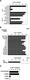
Partial characterization of the HIV-1 suppressor factor. (A) The HIV-1 immune serum and in vitro-restimulated culture supernatants from DC-HIV-1JR-CSF-immune hu-PBL-SCID-spl mice were heated at 56°C for 30 min or separated into heparin-binding andnonbinding fractions by passage of the serum or supernatant fluid through heparin-Sepharose columns (HC). The heparin-bound fraction was eluted with 2 M NaCl buffer. Thereafter, activated PBMC were pretreated with these samples (at final concentrations of 20% serum and 50% culture supernatants) in the absence or presence (+Ab) of a mixture of anti-β-chemokine antibodies and then infected with 500 TCID50 of HIV-1JR-CSF. After 5 days, the p24 level in each culture supernatant was calculated. The percent inhibition was calculated by using the p24 value obtained with the medium control, which was 22.3 ng/ml. (B) Pooled sera (10%) from the DC-HIV-1JR-CSF-immune hu-PBL-SCID-spl mice were preincubated with each anti-human cytokine antibody at 10 μg/ml and then examined for suppressor activity against HIV-1JR-CSF infection of PBMC. In experiment (Exp.) II, the immune serum was pretreated with a mixture of anti-IL-4, anti-IL-13, and anti-IFN-β for 1 h before its addition to activated PBMC, and the PBMC were then infected with HIV-1JR-CSF as described above. The p24 value for the medium control was 20.0 ng/ml. (C) Pooled sera from the DC-HIV-1JR-CSF-immune hu-PBL-SCID-spl mice were passed through the HC column, and then aliquots were filtered through 100-, 50-, and 10-kDa-cutoff Centricon filters. The filtrates obtained were examined for suppressive activity against HIV-1JR-CSF infection of PBMC at a 10% concentration. The data presented are representative of those from four independent experiments. Error bars indicate standard deviations.
FIG. 8.
FIG. 8.
The suppressor factor has no detectable effect on the levels of CCR5 and CD4 expression by macrophages. Macrophages cultured for 5 days with M-CSF were treated with either the immune serum (10%) or the in vitro-restimulated CD4+ T-cell culture supernatants (50%) generated from the DC-HIV-1JR-CSF-immune hu-PBL-SCID mice at 37°C for 1 h. The cells were FcR blocked and stained with anti-CD4 and -CCR5. The data shown are representative of those from four independent experiments. MFI, mean of fluorescence intensity.

Similar articles

Cited by

References

    1. Aaberge, I. S., T. E. Steinsvik, E. C. Groeng, R. B. Leikvold, and M. Lovik. 1996. Human antibody response to a pneumococcal vaccine in SCID-PBL-hu mice and simultaneously vaccinated human cell donors. Clin. Exp. Immunol. 105:12-17. - PMC - PubMed
    1. Adachi, A., H. E. Gendelman, S. Koenig, T. Folks, R. Willey, A. Rabson, and M. A. Martin. 1986. Production of acquired immunodeficiency syndrome-associated retrovirus in human and nonhuman cells transfected with an infectious molecular clone. J. Virol. 59:284-291. - PMC - PubMed
    1. Bombil, F., J. P. Kints, J. M. Scheiff, H. Bazin, and D. Latinne. 1996. A promising model of primary human immunization in human-scid mouse. Immunobiology 195:360-375. - PubMed
    1. Delhem, N., F. Hadida, G. Gorochov, F. Carpentier, J. P. de Cavel, J. F. Andreani, B. Autran, and J. Y. Cesbron. 1998. Primary Th1 cell immunization against HIVgp160 in SCID-hu mice coengrafted with peripheral blood lymphocytes and skin. J. Immunol. 161:2060-2069. - PubMed
    1. Dhawan, S., L. M. Wahl, A. Heredia, Y. Zhang, J. S. Epstein, M. S. Meltzer, and I. K. Hewlett. 1995. Interferon-gamma inhibits HIV-induced invasiveness of monocytes. J. Leukoc. Biol. 58:713-716. - PubMed

Publication types