. 2003 Aug;77(16):8756-64.
doi: 10.1128/jvi.77.16.8756-8764.2003.

Probing the importance of tRNA anticodon: human immunodeficiency virus type 1 (HIV-1) RNA genome complementarity with an HIV-1 that selects tRNA(Glu) for replication

Affiliations

Probing the importance of tRNA anticodon: human immunodeficiency virus type 1 (HIV-1) RNA genome complementarity with an HIV-1 that selects tRNA(Glu) for replication

Lesley C Dupuy et al. J Virol. 2003 Aug.

Abstract

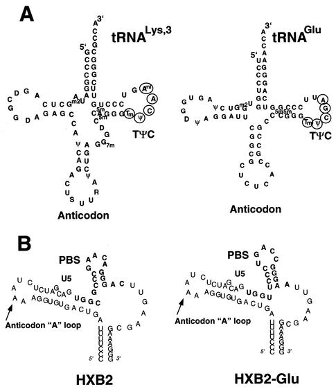
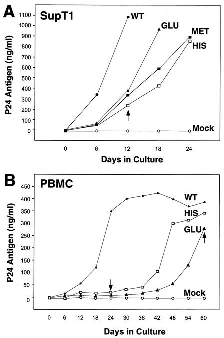
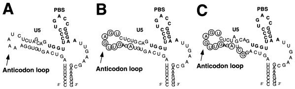
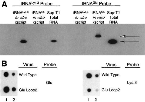
The initiation of human immunodeficiency virus type 1 (HIV-1) reverse transcription occurs at the primer binding site (PBS) that is complementary to the 3'-terminal nucleotides of tRNA(3)(Lys). Why all known strains of HIV-1 select tRNA(3)(Lys) for replication is unknown. Previous studies on the effect of altering the PBS of HIV-1 on replication identified an HIV-1 with a PBS complementary to tRNA(Glu). Since the virus was not initially designed to use tRNA(Glu), the virus had selected tRNA(Glu) from the intracellular pool of tRNA for use in replication. Further characterization of HIV-1 that uses tRNA(Glu) may provide new insights into the preference for tRNA(3)(Lys). HIV-1 constructed with the PBS complementary to tRNA(Glu) was more stable than HIV-1 with the PBS complementary to tRNA(Met) or tRNA(His); however, all of these viruses eventually reverted back to using tRNA(3)(Lys) following growth in SupT1 cells or peripheral blood mononuclear cells (PBMCs). New HIV-1 mutants with nucleotides in U5 complementary to the anticodon of tRNA(Glu) remained stable when grown in SupT1 cells or PBMCs, although the mutants grew more slowly than the wild-type virus. Sequence analysis of the U5 region and the PBS revealed additional mutations predicted to further promote tRNA-viral genome interaction. The results support the importance of the tRNA anticodon-genome interaction in the selection of the tRNA primer and highlight the fact that unique features of tRNA(3)(Lys) are exploited by HIV-1 for selection as the reverse transcription primer.


Figures

FIG. 1.
FIG. 1.
Diagrams of tRNAs and U5-PBS RNA. (A) formula imageand tRNAGlu. The cloverleaf diagrams of formula imageand tRNAGlu are presented with the modified bases in both tRNAs noted. The PBS of HIV is complementary to the 3′-terminal nucleotides of formula image; a second HIV genome was constructed in which the PBS was complementary to the 3′-terminal 18 nucleotides of tRNAGlu. Those nucleotides circled in the TφC-loop of formula imageand tRNAGlu are complementary to regions downstream of the PBS. Abbreviations: S, 5-methoxycarbonylmethyl-2-thiouridine; ψ, pseudouridine; D, dihydrouridine; Tm, 2′-O-methyl-5-methyluridine; Aml, 1-methyladenosine; C5m, 5-methylcytidine; Gm2, N2-methylguanosine; G7m, 7-methylguanosine. (B) The U5-PBS region from HXB2 and that from HXB2(Glu) in which the PBS has been altered to be complementary to tRNAGlu are shown. The PBS is part of a stem-loop structure in which adenines are displayed at the crown of the stem-loop.
FIG. 2.
FIG. 2.
Replication of HIV-1 with altered PBS in SupT1 cells and PBMCs. (A) The replication of the wild-type virus (WT) and viruses with PBSs complementary to tRNAGlu (Glu), tRNAMet (Met), and tRNAHis (His) was analyzed in SupT1 cells. Cultures were initiated as described in Materials and Methods, and the supernatants were analyzed for p24 antigen production during a 24-day culture period. The values represented are from a single experiment; repeat experiments gave similar replication profiles. The arrow denotes the sample time at which PBSs from both viruses which used tRNAMet and tRNAHis reverted to PBSs complementary to formula image(Table 1). (B) Replication of HIV-1 with the PBS complementary to tRNAHis or tRNAGlu in PBMCs. Cultures were initiated as described in Materials and Methods, and virus replication was monitored by analysis of p24 antigen in culture supernatants. The arrow denotes the sample time at which the PBSs complementary to tRNAHis and tRNAGlu were found to revert to PBSs complementary to formula image(Table 2). Results presented are from a single experiment; repeat experiments gave similar replication profiles for each virus.
FIG. 3.
FIG. 3.
HIV-1 with the U5-PBS region complementary to tRNAGlu. HIV-1 genomes in which the PBS and A-loop regions have been altered to be complementary to the 3′-terminal nucleotides and anticodon of tRNAGlu are presented. The PBS region is noted in bold. The circled nucleotides are those which were changed to be complementary to the anticodon of tRNAGlu (CUC). (A) Wild-type HIV-1 genome in which the PBS has been altered to be complementary to tRNAGlu. (B) HIV-1 genome with additional nucleotides making it complementary to the anticodon of tRNAGlu [HXB2(Glu Loop 1)]. An additional nucleotide change was added to enhance the stem of the U5-PBS stem-loop (A, circled). (C) HIV-1 genome in which the PBS and A-loop have been altered to correspond to the 3′-terminal nucleotides of tRNAGlu and the anticodon region. Additional nucleotides inserted within the stem destabilized the stem-loop, creating a bulge region; virus with this genome is designated HXB2(Glu Loop 2).
FIG. 4.
FIG. 4.
Replication of HIV genomes with U5-PBS regions optimized for use of tRNAGlu. (A) Replication in SupT1 cells. Virus cultures were initiated as described in Materials and Methods. The supernatants from the cultures were analyzed for p24 antigen production at various days postinitiation of culture. For comparison, results for viruses in which only the PBS was altered to correspond to tRNAGlu (Glu) are shown. Results for two additional viruses with both the U5 and the PBS altered to correspond to tRNAGlu (Glu-Loop 1 and Glu-Loop 2) are presented. WT, wild type. (B) Replication of viruses in PBMCs. The replication of one of the viruses represented in panel A (Glu-Loop 2) was analyzed in PBMCs. For comparison, results for the virus with only the PBS altered to be complementary to tRNAGlu (Glu) are presented. The arrow denotes the sample time at which the virus with only the PBS complementary to tRNAGlu had a PBS complementary to formula image. Results are from a single experiment; the cultures were performed in duplicate with similar replication profiles.
FIG. 5.
FIG. 5.
Analysis of intracellular and HIV-1 virion-associated formula imageand tRNAGlu. (A) Analysis of tRNA from SupT1 cells. Total RNA from SupT1 cells was isolated as described in Materials and Methods. Following electrophoresis and transfer onto nitrocellulose membrane, equal amounts were probed with oligonucleotides specific for formula imageor tRNAGlu. Specificity controls included equal amounts (1 ng) of in vitro RNA transcription products; note that the in vitro transcript (xscript) for tRNAGlu contains extra nucleotides that retard the migration in the agarose gels used for this analysis. The densities of the probes reacting with the cognate tRNA for both formula imageand tRNAGlu were similar (i.e., the density of the formula imageprobe with 1 ng of in vitro-transcribed formula imageversus the density of the tRNAGlu probe with 1 ng of in vitro-transcribed tRNAGlu). The use of scanning densitometry to determine the cellular amounts of tRNAGlu and formula imagerevealed an approximate 10:1 ratio. This analysis was repeated three times, and a representative blot is shown. The closed arrow denotes migration of full-length tRNALys or tRNAGlu. The open arrow denotes migration of the tRNAGlu in vitro transcript containing additional nucleotides from cloning of the tRNA gene into the transcription plasmid. (B) Analysis of tRNA from HIV-1 virions. Total virion RNA collected from viruses isolated from supernatant of SupT1 cells infected with HXB2 or HXB2(Glu Loop 2). HIV-1 virions were isolated by ultracentrifugation, and RNA was extracted and prepared as described in Materials and Methods. The blot was probed for tRNAGlu and formula imageas indicated. Lane 1, 1 μg of RNA isolated from virions; lane 2, 0.1 μg of RNA isolated from virions.
FIG. 6.
FIG. 6.
Summary of sequence changes found in U5-PBS regions following virus growth in SupT1 cells and PBMCs. (A) Changes found in the U5 region of the virus HXB2(Glu Loop 1) following replication in SupT1 cells. The arrows denote the major changes observed following replication. An additional change of G to A within the A-loop region (circled) was also found. Note that we did not see reversion of the PBS back to the wild-type PBS complementary to formula image. (B) Changes found in the virus HXB2(Glu Loop 2) following in vitro culture in SupT1 cells or PBMCs. The major changes were noted within the stem region of the U5 PBS stem-loop. In this case, C-to-G, G-to-U, and G-to-A changes were noted in viruses grown in SupT1 cells; a G-to-A change was noted in viruses grown in PBMCs and SupT1 cells (PBMC + SupT1). These changes will be predicted to maintain a bulge region reduced from that of the initial HXB2(Glu Loop 2). Note again that reversion of the PBS to the wild-type PBS complementary to formula imagewas found.

Similar articles

Cited by

References

    1. Baltimore, D. 1970. RNA-dependent DNA polymerase in virions of RNA tumour viruses. Nature 226:1209-1211. - PubMed
    1. Beerens, N., B. Klaver, and B. Berkhout. 2000. A structured RNA motif is involved in correct placement of the tRNA3Lys primer onto the human immunodeficiency virus genome. J. Virol. 74:2227-2238. - PMC - PubMed
    1. Berkowitz, R., J. Fisher, and S. P. Goff. 1996. RNA packaging. Curr. Top. Microbiol. Immunol. 214:177-218. - PubMed
    1. Cen, S., A. Khorchid, H. Javanbakht, J. Gabor, T. Stello, K. Shiba, K. Musier-Forsyth, and L. Kleiman. 2001. Incorporation of lysyl-tRNA synthetase into human immunodeficiency virus type 1. J. Virol. 75:5043-5048. - PMC - PubMed
    1. Cimarelli, A., and J. Luban. 1999. Translation elongation factor 1-alpha interacts specifically with the human immunodeficiency virus type 1 Gag polyprotein. J. Virol. 73:5388-5401. - PMC - PubMed

Publication types

LinkOut - more resources