

Development of recombinant vesicular stomatitis viruses that exploit defects in host defense to augment specific oncolytic activity
- PMID: 12885903
- PMCID: PMC167243
- DOI: 10.1128/jvi.77.16.8843-8856.2003
Development of recombinant vesicular stomatitis viruses that exploit defects in host defense to augment specific oncolytic activity
Abstract
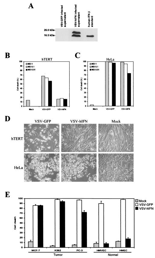
Vesicular stomatitis virus (VSV) is a negative-stranded RNA virus normally sensitive to the antiviral actions of alpha/beta interferon (IFN-alpha/beta). Recently, we reported that VSV replicates to high levels in many transformed cells due, in part, to susceptible cells harboring defects in the IFN system. These observations were exploited to demonstrate that VSV can be used as a viral oncolytic agent to eradicate malignant cells in vivo while leaving normal tissue relatively unaffected. To attempt to improve the specificity and efficacy of this system as a potential tool in gene therapy and against malignant disease, we have genetically engineered VSV that expresses the murine IFN-beta gene. The resultant virus (VSV-IFNbeta) was successfully propagated in cells not receptive to murine IFN-alpha/beta and expressed high levels of functional heterologous IFN-beta. In normal murine embryonic fibroblasts (MEFs), the growth of VSV-IFNbeta was greatly reduced and diminished cytopathic effect was observed due to the production of recombinant IFN-beta, which by functioning in a manner involving autocrine and paracrine mechanisms induced an antiviral effect, preventing virus growth. However, VSV-IFNbeta grew to high levels and induced the rapid apoptosis of transformed cells due to defective IFN pathways being prevalent and thus unable to initiate proficient IFN-mediated host defense. Importantly, VSV expressing the human IFN-beta gene (VSV-hIFNbeta) behaved comparably and, while nonlytic to normal human cells, readily killed their malignant counterparts. Similar to our in vitro observations, following intravenous and intranasal inoculation in mice, recombinant VSV (rVSV)-IFNbeta was also significantly attenuated compared to wild-type VSV or rVSV expressing green fluorescent protein. However, VSV-IFNbeta retained propitious oncolytic activity against metastatic lung disease in immunocompetent animals and was able to generate robust antitumor T-cell responses. Our data indicate that rVSV designed to exploit defects in mechanisms of host defense can provide the basis for new generations of effective, specific, and safer viral vectors for the treatment of malignant and other disease.
Figures
Similar articles
-
Genetically engineered vesicular stomatitis virus in gene therapy: application for treatment of malignant disease.J Virol. 2002 Jan;76(2):895-904. doi: 10.1128/jvi.76.2.895-904.2002. J Virol. 2002. PMID: 11752178 Free PMC article.
-
Potent systemic therapy of multiple myeloma utilizing oncolytic vesicular stomatitis virus coding for interferon-β.Cancer Gene Ther. 2012 Jul;19(7):443-50. doi: 10.1038/cgt.2012.14. Epub 2012 Apr 20. Cancer Gene Ther. 2012. PMID: 22522623 Free PMC article.
-
Vesicular stomatitis virus expressing interferon-β is oncolytic and promotes antitumor immune responses in a syngeneic murine model of non-small cell lung cancer.Oncotarget. 2015 Oct 20;6(32):33165-77. doi: 10.18632/oncotarget.5320. Oncotarget. 2015. PMID: 26431376 Free PMC article.
-
Vesicular stomatitis virus as an oncolytic vector.Viral Immunol. 2004;17(4):516-27. doi: 10.1089/vim.2004.17.516. Viral Immunol. 2004. PMID: 15671748 Review.
-
VSV-tumor selective replication and protein translation.Oncogene. 2005 Nov 21;24(52):7710-9. doi: 10.1038/sj.onc.1209042. Oncogene. 2005. PMID: 16299531 Review.
Cited by
-
STAT3 inhibition reduces toxicity of oncolytic VSV and provides a potentially synergistic combination therapy for hepatocellular carcinoma.Cancer Gene Ther. 2015 Jun;22(6):317-25. doi: 10.1038/cgt.2015.23. Epub 2015 May 1. Cancer Gene Ther. 2015. PMID: 25930184
-
PEGylation of vesicular stomatitis virus extends virus persistence in blood circulation of passively immunized mice.J Virol. 2013 Apr;87(7):3752-9. doi: 10.1128/JVI.02832-12. Epub 2013 Jan 16. J Virol. 2013. PMID: 23325695 Free PMC article.
-
Efficient Delivery and Replication of Oncolytic Virus for Successful Treatment of Head and Neck Cancer.Int J Mol Sci. 2020 Sep 25;21(19):7073. doi: 10.3390/ijms21197073. Int J Mol Sci. 2020. PMID: 32992948 Free PMC article. Review.
-
Vesicular stomatitis virus variants selectively infect and kill human melanomas but not normal melanocytes.J Virol. 2013 Jun;87(12):6644-59. doi: 10.1128/JVI.03311-12. Epub 2013 Apr 3. J Virol. 2013. PMID: 23552414 Free PMC article.
-
Characterization of resistance to rhabdovirus and retrovirus infection in a human myeloid cell line.PLoS One. 2015 Mar 26;10(3):e0121455. doi: 10.1371/journal.pone.0121455. eCollection 2015. PLoS One. 2015. PMID: 25811758 Free PMC article.
References
-
- Adkins, B., Y. Bu, and P. Guevara. 2001. The generation of Th memory in neonates versus adults: prolonged primary Th2 effector function and impaired development of Th1 memory effector function in murine neonates. J. Immunol. 166:918-925. - PubMed
-
- Albini, A., C. Marchisone, F. Del Grosso, R. Benelli, L. Masiello, C. Tacchetti, M. Bono, M. Ferrantini, C. Rozera, M. Truini, F. Belardelli, L. Santi, and D. M. Noonan. 2000. Inhibition of angiogenesis and vascular tumor growth by interferon-producing cells: a gene therapy approach. Am. J. Pathol. 156:1381-1393. - PMC - PubMed
-
- Balachandran, S., and G. N. Barber. 2000. Vesicular stomatitis virus therapy of tumors. IUBMB Life 50:1-4. - PubMed
MeSH terms
Substances
LinkOut - more resources
Full Text Sources
Other Literature Sources
