Deletions in the putative cell receptor-binding domain of Sindbis virus strain MRE16 E2 glycoprotein reduce midgut infectivity in Aedes aegypti
- PMID: 12885905
- PMCID: PMC167217
- DOI: 10.1128/jvi.77.16.8872-8881.2003
Deletions in the putative cell receptor-binding domain of Sindbis virus strain MRE16 E2 glycoprotein reduce midgut infectivity in Aedes aegypti
Abstract
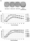
The Sindbis virus (Alphavirus; Togaviridae) strain MRE16 efficiently infects Aedes aegypti mosquitoes that ingest a blood meal containing 8 to 9 log(10) PFU of virus/ml. However, a small-plaque variant of this virus, MRE16sp, poorly infects mosquitoes after oral infection with an equivalent titer. To determine the genetic differences between MRE16 and MRE16sp viruses, we have sequenced the MRE16sp structural genes and found a 90-nucleotide deletion in the E2 glycoprotein that spans the 3' end of the coding region for the putative cell-receptor binding domain (CRBD). We examined the role of this deletion in oral infection of mosquitoes by constructing infectious clones pMRE16icDeltaE200-Y229 and pMRE16ic, representing MRE16 virus genomes with and without the deletion, respectively. A third infectious clone, pMRE16icDeltaE200-C220, was also constructed that contained a smaller deletion extending only to the 3' terminus of the CRBD coding region. Virus derived from pMRE16ic replicated with the same efficiency as parental virus in vertebrate (BHK-21) and mosquito (C6/36) cells and orally infected A. aegypti. Viruses derived from pMRE16icDeltaE200-Y229 and pMRE16icDeltaE200-C220 replicated 10- to 100-fold less efficiently in C6/36 and BHK-21 cells than did MRE16ic virus. Each deletion mutant poorly infected A. aegypti and dramatically reduced midgut infectivity and dissemination. However, all viruses generated nearly equal titers (approximately 6.0 log(10) PFU/ml) in mosquitoes 4 days after infection by intrathoracic inoculation. These results suggest that the deleted portion of the E2 CRBD represents an important determinant of MRE16 virus midgut infectivity in A. aegypti.
Figures


Similar articles
-
Genetic determinants of Sindbis virus mosquito infection are associated with a highly conserved alphavirus and flavivirus envelope sequence.J Virol. 2008 Mar;82(6):2966-74. doi: 10.1128/JVI.02060-07. Epub 2007 Dec 26. J Virol. 2008. PMID: 18160430 Free PMC article.
-
Development of a chimeric sindbis virus with enhanced per Os infection of Aedes aegypti.Virology. 1998 Mar 30;243(1):99-112. doi: 10.1006/viro.1998.9034. Virology. 1998. PMID: 9527919
-
Infection of Aedes aegypti Mosquitoes with Midgut-Attenuated Sindbis Virus Reduces, but Does Not Eliminate, Disseminated Infection.J Virol. 2021 Jun 10;95(13):e0013621. doi: 10.1128/JVI.00136-21. Epub 2021 Jun 10. J Virol. 2021. PMID: 33853958 Free PMC article.
-
Genetic determinants of Sindbis virus strain TR339 affecting midgut infection in the mosquito Aedes aegypti.J Gen Virol. 2007 May;88(Pt 5):1545-1554. doi: 10.1099/vir.0.82577-0. J Gen Virol. 2007. PMID: 17412985
-
Comparison of the transmission potential of two genetically distinct Sindbis viruses after oral infection of Aedes aegypti (Diptera: Culicidae).J Med Entomol. 2004 Jan;41(1):95-106. doi: 10.1603/0022-2585-41.1.95. J Med Entomol. 2004. PMID: 14989352
Cited by
-
Sequential adaptive mutations enhance efficient vector switching by Chikungunya virus and its epidemic emergence.PLoS Pathog. 2011 Dec;7(12):e1002412. doi: 10.1371/journal.ppat.1002412. Epub 2011 Dec 8. PLoS Pathog. 2011. PMID: 22174678 Free PMC article.
-
Role of phosphatidylserine receptors in enveloped virus infection.J Virol. 2014 Apr;88(8):4275-90. doi: 10.1128/JVI.03287-13. Epub 2014 Jan 29. J Virol. 2014. PMID: 24478428 Free PMC article.
-
Evolutionary ecology of virus emergence.Ann N Y Acad Sci. 2017 Feb;1389(1):124-146. doi: 10.1111/nyas.13304. Epub 2016 Dec 30. Ann N Y Acad Sci. 2017. PMID: 28036113 Free PMC article. Review.
-
Stress granule components G3BP1 and G3BP2 play a proviral role early in Chikungunya virus replication.J Virol. 2015 Apr;89(8):4457-69. doi: 10.1128/JVI.03612-14. Epub 2015 Feb 4. J Virol. 2015. PMID: 25653451 Free PMC article.
-
Differential transmission of Asian and African Zika virus lineages by Aedes aegypti from New Caledonia.Emerg Microbes Infect. 2018 Sep 26;7(1):159. doi: 10.1038/s41426-018-0166-2. Emerg Microbes Infect. 2018. PMID: 30254274 Free PMC article.
References
-
- Arcus, Y. M., E. J. Houk, and J. L. Hardy. 1983. Comparative in vitro binding of an arbovirus to midgut microvillar membranes from susceptible and refractory Culex mosquitoes. Fed. Proc. 42:2141.
-
- Burness, A. T., I. Pardoe, S. G. Faragher, S. Vrati, and L. Dalgarno. 1988. Genetic stability of Ross River virus during epidemic spread in nonimmune humans. Virology 167:639-643. - PubMed
-
- Chanas, A. C., E. A. Gould, J. C. Clegg, and M. G. Varma. 1982. Monoclonal antibodies to Sindbis virus glycoprotein E1 can neutralize, enhance infectivity, and independently inhibit haemagglutination or haemolysis. J. Gen. Virol. 58:37-46. - PubMed
-
- Davis, N. L., D. F. Pence, W. J. Meyer, A. L. Schmaljohn, and R. E. Johnston. 1987. Alternative forms of a strain-specific neutralizing antigenic site on the Sindbis virus E2 glycoprotein. Virology 161:101-108. - PubMed
Publication types
MeSH terms
Substances
Grants and funding
LinkOut - more resources
Full Text Sources
Other Literature Sources
Molecular Biology Databases

