. 2003 Aug;77(16):8924-33.
doi: 10.1128/jvi.77.16.8924-8933.2003.

Incorporation of tick-borne encephalitis virus replicons into virus-like particles by a packaging cell line

Affiliations

Incorporation of tick-borne encephalitis virus replicons into virus-like particles by a packaging cell line

Rainer Gehrke et al. J Virol. 2003 Aug.

Abstract

RNA replicons derived from flavivirus genomes show considerable potential as gene transfer and immunization vectors. A convenient and efficient encapsidation system is an important prerequisite for the practical application of such vectors. In this work, tick-borne encephalitis (TBE) virus replicons and an appropriate packaging cell line were constructed and characterized. A stable CHO cell line constitutively expressing the two surface proteins prM/M and E (named CHO-ME cells) was generated and shown to efficiently export mature recombinant subviral particles (RSPs). When replicon NdDeltaME lacking the prM/M and E genes was introduced into CHO-ME cells, virus-like particles (VLPs) capable of initiating a single round of infection were released, yielding titers of up to 5 x 10(7)/ml in the supernatant of these cells. Another replicon (NdDeltaCME) lacking the region encoding most of the capsid protein C in addition to proteins prM/M and E was not packaged by CHO-ME cells. As observed with other flavivirus replicons, both TBE virus replicons appeared to exert no cytopathic effect on their host cells. Sedimentation analysis revealed that the NdDeltaME-containing VLPs were physically distinct from RSPs and similar to infectious virions. VLPs could be repeatedly passaged in CHO-ME cells but maintained the property of being able to initiate only a single round of infection in other cells during these passages. CHO-ME cells can thus be used both as a source for mature TBE virus RSPs and as a safe and convenient replicon packaging cell line, providing the TBE virus surface proteins prM/M and E in trans.


Figures

FIG. 1.
FIG. 1.
Structure of TBE virus replicons. (A) Schematic of the TBE virus genome (top) and the locations of the introduced deletions (below, double-headed arrows together with the corresponding plasmid designations). The open reading frame is represented by an open box, with only the structural protein coding region drawn to scale. Internal signal sequences are shown as black bars. Positions of cleavage sites of restriction enzymes used during cloning (nucleotide numbers refer to the wild-type genome of TBE virus) and amino acid residues flanking the deletions (numbering starts with the first amino acid residue of protein C or protein E) are indicated above and below the schematic, respectively. NCR, noncoding region. (B) Sequence details of the junction regions. Nucleotide and amino acid residues of the wild-type TBE virus sequence flanking the deletions are marked above and below the sequences, respectively. Recognition sites for restriction enzymes are underlined (MluI, ACG CGT; BamHI, GGA TCC). In the case of plasmid pTNdΔCME, 15 nucleotides coding for an artificial NS2B/3 protease cleavage site and creating the BamHI restriction site were introduced. The insertion of a T residue in plasmid pTNdΔME created a BamHI cleavage site (underlined) and maintained the authentic residue 100 (Asp) of protein C. Arrows depict the sites at which the amino acid sequences are presumed to be cleaved by the viral protease NS2B/3.
FIG. 2.
FIG. 2.
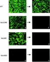
Protein expression (left column) and attempted cell culture passage (right column) of TBE virus replicons. Wild-type (WT, A), NdΔCME (B), NdΔME (C), and replication-deficient mutant NdΔNS5 (D) RNAs were synthesized in vitro and transfected into BHK-21 cells. Expression of protein NS1 was tested 72 h posttransfection by immunofluorescence. (E to H) Supernatants were passaged (as indicated by arrows) onto fresh BHK-21 cell cultures, and the expression of protein NS1 was determined 72 h postinoculation by immunofluorescence.
FIG. 3.
FIG. 3.
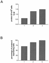
Monitoring of protein E release (A) and proportion of protein E-producing cells (B) during the derivation of CHO-ME cells. Protein E was quantified 72 h postseeding of cells by SDS-ELISA. Protein E-producing cells were identified by immunofluorescence staining. 1, cells derived from a single-cell colony after transfection of CHO-DG44 cells with the expression plasmids; 2, cells after the first subcloning step; 3, CHO-ME cells as derived after a second subcloning step.
FIG. 4.
FIG. 4.
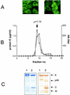
Characterization of CHO-ME cells and released particles. (A) Cell morphology and protein E expression of low-passage cells (left) and after 15 passages (right) as examined by immunofluorescence. (B) Determination of buoyant density of secreted particles. Particles were precipitated from supernatants of CHO-ME cells purified by rate zonal centrifugation and then subjected to sucrose density gradient centrifugation. Individual fractions were tested for their protein E content by SDS-ELISA (open circles) and the presence of hemagglutination activity (open boxes). The buoyant density, as determined for the peak fraction, is indicated above the graph. (C) Protein analysis of purified particles by fractionation on a 15% denaturing polyacrylamide gel. Proteins were visualized by Coomassie blue staining (left) or immunoblot analysis (right). 1, CHO-ME derived particles; 2, wild-type virus. The positions of viral structural proteins are indicated on the right.
FIG. 5.
FIG. 5.
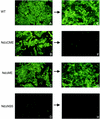
Formation of infectious particles in CHO-ME cells transfected with in vitro-synthesized wild-type (A), NdΔCME (B), NdΔME (C), and NdΔNS5 (D) RNAs. Expression of protein NS1 was visualized by immunofluorescence 72 h posttransfection. (E to H) Supernatants were passaged onto fresh Vero cell cultures as indicated by the arrows, and the expression of protein NS1 was determined 96 h postinoculation.
FIG. 6.
FIG. 6.
Sedimentation analysis of particles harvested from supernatants of CHO-ME cells transfected with in vitro-synthesized NdΔME replicon RNA. Particles were precipitated with polyethylene glycol and applied to a 10 to 50% sucrose gradient. The protein E concentration of individual fractions (open circles) was determined by SDS-ELISA. The presence of replicon RNA in individual fractions was assessed by a semiquantitative RT-PCR and is shown as a percentage of the total from all of the fractions (open diamonds). Control gradients loaded with purified preparations of RSPs and wild-type TBE virus were analyzed in parallel (peak fractions indicated by arrows).

Similar articles

Cited by

References

    1. Aberle, J. H., S. W. Aberle, S. L. Allison, K. Stiasny, M. Ecker, C. W. Mandl, R. Berger, and F. X. Heinz. 1999. A DNA immunization model study with constructs expressing the tick-borne encephalitis virus envelope protein E in different physical forms. J. Immunol. 163:6756-6761. - PubMed
    1. Allison, S. L., C. W. Mandl, C. Kunz, and F. X. Heinz. 1994. Expression of cloned envelope protein genes from the flavivirus tick-borne encephalitis virus in mammalian cells and random mutagenesis by PCR. Virus Genes 8:187-198. - PubMed
    1. Allison, S. L., K. Stadler, C. W. Mandl, C. Kunz, and F. X. Heinz. 1995. Synthesis and secretion of recombinant tick-borne encephalitis virus protein E in soluble and particulate form. J. Virol. 69:5816-5820. - PMC - PubMed
    1. Blight, K. J., A. A. Kolykhalov, and C. M. Rice. 2000. Efficient initiation of HCV RNA replication in cell culture. Science 290:1972-1974. - PubMed
    1. Bredenbeek, P. J., I. Frolov, C. M. Rice, and S. Schlesinger. 1993. Sindbis virus expression vectors: packaging of RNA replicons by using defective helper RNAs. J. Virol. 67:6439-6446. - PMC - PubMed

Publication types