. 2003 Aug;77(16):8973-84.
doi: 10.1128/jvi.77.16.8973-8984.2003.

Complete genomic sequencing shows that polioviruses and members of human enterovirus species C are closely related in the noncapsid coding region

Affiliations

Complete genomic sequencing shows that polioviruses and members of human enterovirus species C are closely related in the noncapsid coding region

Betty Brown et al. J Virol. 2003 Aug.

Abstract

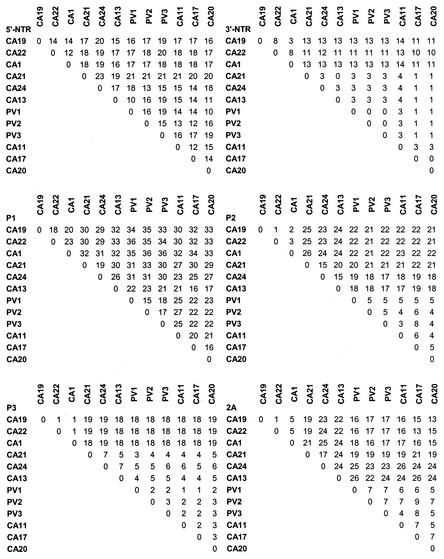
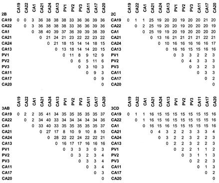
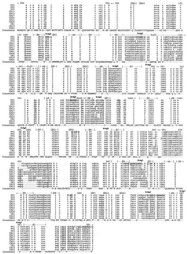
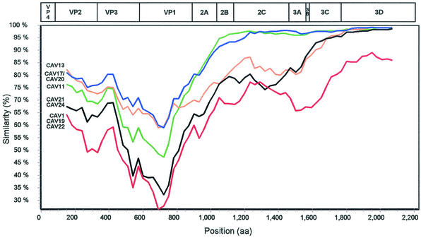
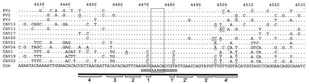
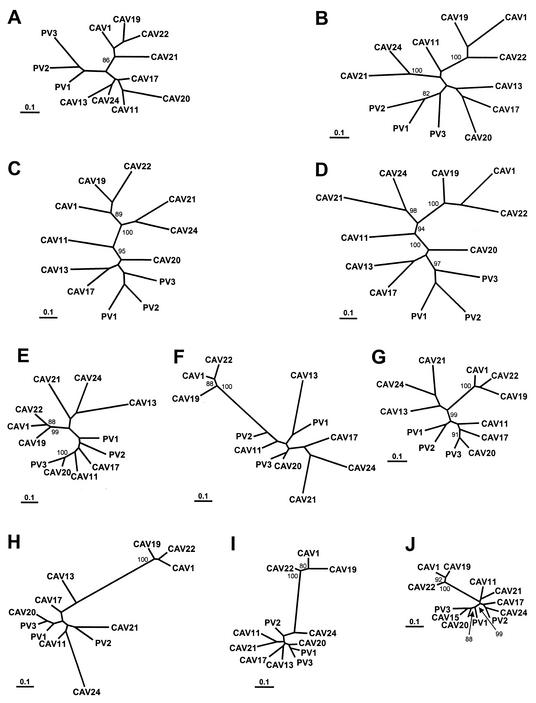
The 65 human enterovirus serotypes are currently classified into five species: Poliovirus (3 serotypes), Human enterovirus A (HEV-A) (12 serotypes), HEV-B (37 serotypes), HEV-C (11 serotypes), and HEV-D (2 serotypes). Coxsackie A virus (CAV) serotypes 1, 11, 13, 15, 17, 18, 19, 20, 21, 22, and 24 constitute HEV-C. We have determined the complete genome sequences for the remaining nine HEV-C serotypes and compared them with the complete sequences of CAV21, CAV24, and the polioviruses. The viruses were most diverse in the capsid region (4 to 36% amino acid difference). A high degree of capsid sequence conservation (96% amino acid identity) suggests that CAV15 and CAV18 should be classified as strains of CAV11 and CAV13, respectively. In the 3CD region, CAV1, CAV19, and CAV22 differed from one another by only 1.2 to 1.4% and CAV11, CAV13, CAV17, CAV20, CAV21, CAV24, and the polioviruses differed from one another by only 1.2 to 3.6%. The two groups, however, differed from one another by 14.6 to 16.2%. The polioviruses as a group were monophyletic only in the capsid region. Only one group of serotypes (CAV1, CAV19, and CAV22) was consistently monophyletic in multiple genome regions. Incongruities among phylogenetic trees based on different genome regions strongly suggest that recombination has occurred between the polioviruses, CAV11, CAV13, CAV17, and CAV20. The close relationship among the polioviruses and CAV11, CAV13, CAV17, CAV20, CAV21, and CAV24 and the uniqueness of CAV1, CAV19, and CAV22 suggest that revisions should be made to the classification of these viruses.


Figures

FIG. 1.
FIG. 1.
Nucleotide and deduced amino acid sequence relationships (percent identity). For noncoding regions (5′NTR and 3′-NTR), nucleotide comparisons are shown. Deduced amino acid sequence comparisons are shown for all other regions.
FIG. 1.
FIG. 1.
Nucleotide and deduced amino acid sequence relationships (percent identity). For noncoding regions (5′NTR and 3′-NTR), nucleotide comparisons are shown. Deduced amino acid sequence comparisons are shown for all other regions.
FIG. 2.
FIG. 2.
Aligned poliovirus and HEV-C capsid precursor proteins. Cleavage sites are indicated above the alignment. Regions predicted to form structurally defined alpha helices and beta strands (16, 29, 36), as well as the location of neutralization antigenic sites N-Ag1, N-Ag2, and N-Ag3 in PV1 (9), are also indicated. Amino acids implicated by structural or genetic studies to be involved in PV1-poliovirus receptor interaction are shaded (2, 15).
FIG. 3.
FIG. 3.
Similarity plots of poliovirus and HEV-C viruses deduced polyprotein sequences, calculated and plotted by SimPlot 3.2 (24), relative to the polyprotein consensus sequence of PV1, PV2, and PV3. To facilitate the analysis, CAV1, CAV19, and CAV22 were grouped together, as were CAV17 and CAV20 and also CAV21 and CAV24, as described in the text. Each point represents the percent identity to the poliovirus consensus, within a sliding window of 300 amino acids centered on the position plotted, with a step of 30 amino acids between points. Positions containing gaps were excluded from the analysis. The PV1 genetic map is shown at the top, approximately to scale.
FIG. 4.
FIG. 4.
Nucleotide sequence alignment of the region surrounding the poliovirus cis-acting replication element (cre). The majority rule consensus sequence is shown at the bottom (Con). Residues that are identical to those of the consensus are indicated by a dot. The bar below the sequence alignment indicates the location of the conserved stem-loop structure. The conserved AAACA motif in the loop is boxed (12, 41), and the location of the RN3AARN6R motif is indicated below the consensus (52). Base-paired residues in the stem are indicated by brackets and numbered 1-1′ to 4-4′, as previously described (12). Residues predicted to disrupt base pairing are underlined. Nucleotides are numbered according to the genome of PV1-Mahoney.
FIG. 5.
FIG. 5.
Phylogenetic trees based on poliovirus and HEV-C virus nucleotide sequences. Each of the major functional regions of the genome was analyzed independently. Bootstrap values (percent of 100 pseudoreplicate data sets) of over 80% supporting each cluster are shown at the nodes. All trees are plotted to the same scale (see scale bar). (A) 5′-NTR; (B) 3′-NTR; (C) complete P1 region; (D) complete P2 region; (E) complete P3 region.
FIG. 6.
FIG. 6.
Phylogenetic trees based on poliovirus and HEV-C virus nucleotide sequences. The regions encoding each of the mature viral proteins were analyzed independently. Bootstrap values (percent of 100 pseudoreplicate data sets) of over 80% supporting each cluster are shown at the nodes. All trees are plotted to the same scale (see scale bar). (A) 1A (VP4); (B) 1B (VP2); (C) 1C (VP3); (D) 1D (VP1); (E) 2A; (F) 2B; (G) 2C; (H) 3AB; (I) 3C; (J) 3D.

References

    1. Andersson, P., K. Edman, and A. M. Lindberg. 2002. Molecular analysis of the echovirus 18 prototype. Evidence of interserotypic recombination with echovirus 9. Virus Res. 85:71-83. - PubMed
    1. Belnap, D. M., B. M. J. McDermott, D. J. Filman, N. Cheng, B. L. Trus, H. J. Zuccola, V. R. Racaniello, J. M. Hogle, and A. C. Steven. 2000. Three-dimensional structure of poliovirus receptor bound to poliovirus. Proc. Natl. Acad. Sci. USA 97:73-78. - PMC - PubMed
    1. Cammack, N., A. Phillips, G. Dunn, V. Patel, and P. D. Minor. 1988. Intertypic genomic rearrangements of poliovirus strains in vaccinees. Virology 167:507-514. - PubMed
    1. Caro, V., S. Guillot, F. Delpeyroux, and R. Crainic. 2001. Molecular strategy for ′serotyping' of human enteroviruses. J. Gen. Virol. 82:79-91. - PubMed
    1. Casas, I., G. F. Palacios, G. Trallero, D. Cisterna, M. C. Freire, and A. Tenorio. 2001. Molecular characterization of human enteroviruses in clinical samples: comparison between VP2, VP1, and RNA polymerase regions using RT nested PCR assays and direct sequencing of products. J. Med. Virol. 65:138-148. - PubMed

LinkOut - more resources