

doi: 10.1128/jvi.77.16.9084-9089.2003.
Recombinant avian infectious bronchitis virus expressing a heterologous spike gene demonstrates that the spike protein is a determinant of cell tropism
Affiliations
- PMID: 12885925
- PMCID: PMC167237
- DOI: 10.1128/jvi.77.16.9084-9089.2003
Item in Clipboard
Recombinant avian infectious bronchitis virus expressing a heterologous spike gene demonstrates that the spike protein is a determinant of cell tropism
J Virol.
2003 Aug.
Abstract
A recombinant infectious bronchitis virus (IBV), BeauR-M41(S), was generated using our reverse genetics system (R. Casais, V. Thiel, S. G. Siddell, D. Cavanagh, and P. Britton, J. Virol. 75:12359-12369, 2001), in which the ectodomain region of the spike gene from IBV M41-CK replaced the corresponding region of the IBV Beaudette genome. BeauR-M41(S) acquired the same cell tropism phenotype as IBV M41-CK in four different cell types, demonstrating that the IBV spike glycoprotein is a determinant of cell tropism.
Figures
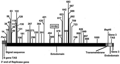
Schematic diagram of the IBV S gene. The 5′ end of the S gene overlaps the 3′ end of the replicase gene. The four domains of the S protein, the position of the S1/S2 cleavage point, and the positions of the PacI and BspHI restriction sites, gene 3 TAS, and the start of gene 3 are shown. The numbers refer to the positions of the amino acid differences resulting from nonsynonymous substitutions following exchange of the two S gene sequences.
Schematic diagram for the construction of the chimeric S gene and production of a full-length IBV cDNA. (A) Replacement of the ectodomain region of the Beaudette S gene by the corresponding sequence from IBV M41 for construction of FRAG-3-M41S. (B) Junction of the S gene sequences at the PacI and BspHI restriction sites with the intervening sequence derived from M41. The nucleotides in bold correspond to the signal and transmembrane sequences, respectively. (C) Schematic diagram of the BeauR-M41(S) full-length cDNA composed of FRAG-1, FRAG-2, and FRAG-3-M41S.
Growth profiles of the three IBVs on four cell types. Cells were infected with 1.5 × 10⁶ PFU of each IBV, and cell medium was analyzed for progeny virus by plaque titration assay on CK cells 0 to 96 h postinfection. The panels show the growth patterns of Beau-R (solid line with triangle), M41-CK (dashed line with diamond), and BeauR-M41(S) (dotted line with square) on CK cells (A), Vero cells (B), CEF cells (C), and BHK-21 cells (D).
Detection of IBV by RT-PCR following passage of Beau-R, M41-CK, or BeauR-M41(S) on CK, Vero, CEF, or BHK-21 cells. Cells (P1) were infected with 2 × 10⁷ PFU of each virus, and 24 h postinfection any progeny viruses were passaged twice (P1 to P3). Total cellular RNA was isolated from the IBV-infected cells and used for RT-PCR analysis; an RT-PCR product of 666 bp corresponded to detection of Beau-R- and BeauR-M41(S)- derived RNA, and a product of 481 bp corresponded to detection of M41-CK-derived RNA. The RT-PCR products detected following passage (P1 to P3) of Beau-R, M41-CK, or BeauR-M41(S) on CK (A), Vero (B), CEF (C), or BHK-21 (D) cells were analyzed on 1% agarose gels.
References
-
- Cavanagh, D. 2001. A nomenclature for avian coronavirus isolates and the question of species status. Avian Pathol. 30:109-115. - PubMed
-
- Cavanagh, D., K. Mawditt, M. Sharma, S. E. Drury, H. L. Ainsworth, P. Britton, and R. E. Gough. 2001. Detection of a coronavirus from turkey poults in Europe genetically related to infectious bronchitis virus of chickens. Avian Pathol. 30:365-378. - PubMed
-
- Cavanagh, D., K. Mawditt, D. B. B. Welchman, P. Britton, and R. E. Gough. 2002. Coronaviruses from pheasants (Phasianus colchicus) are genetically closely related to coronaviruses of domestic fowl (infectious bronchitis virus) and turkeys. Avian Pathol. 31:81-93. - PubMed
Publication types
MeSH terms
Substances
LinkOut - more resources
Full Text Sources
Other Literature Sources
Research Materials
