. 2003 Aug 19;100(17):9861-6.
doi: 10.1073/pnas.1633363100. Epub 2003 Jul 28.

A high-molecular-weight complex of membrane proteins BAP29/BAP31 is involved in the retention of membrane-bound IgD in the endoplasmic reticulum

Affiliations

A high-molecular-weight complex of membrane proteins BAP29/BAP31 is involved in the retention of membrane-bound IgD in the endoplasmic reticulum

Wolfgang W A Schamel et al. Proc Natl Acad Sci U S A. .

Abstract

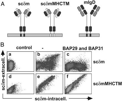
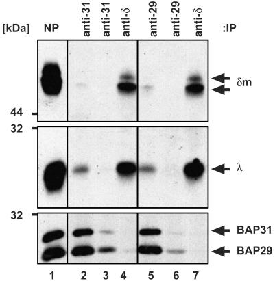
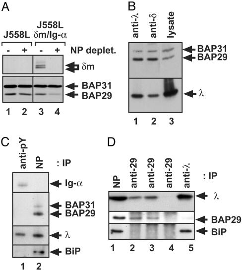
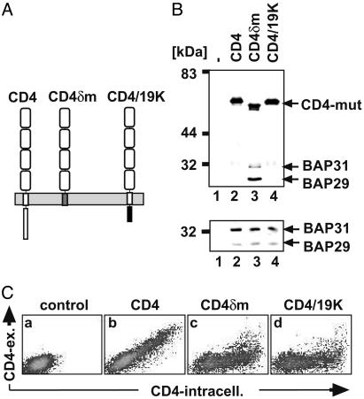
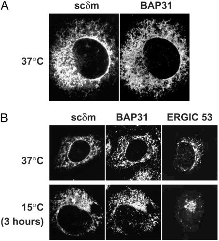
B cell antigen receptors (BCRs) are multimeric transmembrane protein complexes comprising membrane-bound immunoglobulins (mIgs) and Ig-alpha/Ig-beta heterodimers. In most cases, transport of mIgs from the endoplasmic reticulum (ER) to the cell surface requires assembly with the Ig-alpha/Ig-beta subunits. In addition to Ig-alpha/Ig-beta, mIg molecules also bind two ER-resident membrane proteins, BAP29 and BAP31, and the chaperone heavy chain binding protein (BiP). In this article, we show that neither Ig-alpha/Ig-beta nor BAP29/BAP31 nor BiP bind simultaneously to the same mIgD molecule. Blue native PAGE revealed that only a minor fraction of intracellular mIgD is associated with high-molecular-weight BAP29/BAP31 complexes. BAP-binding to mIgs was found to correlate with ER retention of chimeric mIgD molecules. On high-level expression in Drosophila melanogaster S2 cells, mIgD molecules were detected on the cell surface in the absence of Ig-alpha/Ig-beta. This aberrant transport was prevented by coexpression of BAP29 and BAP31. Thus, BAP complexes contribute to ER retention of mIg complexes that are not bound to Ig-alpha/Ig-beta. Furthermore, the mechanism of ER retention of both BAP31 and mIgD is not through retrieval from a post-ER compartment, but true ER retention. In conclusion, BAP29 and BAP31 might be the long sought after retention proteins and/or chaperones that act on transmembrane regions of various proteins.


Figures

Fig. 5.
Fig. 5.
Expression of scδm molecules on the cell surface. (A) Images of scδm, scδmMHCTM, and mIgD molecules are shown. The heavy chain-derived sequence is dark gray, the light chain-derived sequence is light gray, and the TM region of MHCI is white. (B) Untransfected S2 cells (Ba and Bd)orscδm(b) and scδmMHCTM (e) transfectants, as well as BAP29/BAP31/scδm (c) and BAP29/BAP31/scδmMHCTM cotransfectants (f), were stained first on the cell surface (phycoerythrin) and then intracellularly (FITC) with an anti-sc antibody. Cells were analyzed as shown in Fig. 4.
Fig. 1.
Fig. 1.
Triton X-100-solubilized mIgD complexes exclusively contain either Ig-α/Ig-β or BAP29/BAP31. Phosphotyrosine-containing proteins were purified from stimulated J558Lδm/Ig-α cells (lanes 1), and mIgD complexes remaining in the supernatant were purified with NP-Sepharose (lanes 2). Proteins were separated by SDS/PAGE and detected with anti-phosphotyrosine antibodies (A) and anti-δ, anti-Ig-β, and a mixture of anti-BAP29 and anti-BAP31 antibodies (B).
Fig. 2.
Fig. 2.
Only minor amounts of mIgD are associated with BAP29/BAP31. The mIgD complexes were purified from J558Lδm/Ig-α lysates with NP-Sepharose (lane1), eluted by free hapten, and precipitated twice with either an anti-BAP31 (lanes 2 and 3) or an anti-BAP29 antiserum (lanes 5 and 6). Subsequently, the mIgD molecules remaining in the supernatants were purified with an anti-δ antiserum (lanes 4 and 7). Proteins were separated by SDS/PAGE and detected with anti-δ, anti-λ, anti-BAP29, and anti-BAP31 antisera.
Fig. 3.
Fig. 3.
BAP31 occurs in a homomeric BAP31 and in a heterodimeric BAP29/BAP31 pool; only the latter is associated with mIgD. (A) Western blot analysis of δm(Upper) and BAP29/BAP31 (Lower) in cell lysates of J558L (lane 1 and 2) and J558Lδm/Ig-α cells (lane 3 and 4). The lysates were left untreated (lane 1 and 3) or incubated with NP-Sepharose to remove mIgD from the lysates (lane 2 and 4). (B) mIgD complexes were purified from J558Lδm/Ig-α lysates with anti-λ (lane 1) or anti-δ (lane 2) antisera. Proteins were separated by SDS/PAGE and detected with anti-BAP29, anti-BAP31, and anti-λ antisera. For comparison, a cellular lysate was used (lane 3). (C) Phosphotyrosine-containing proteins were purified from stimulated J558Lδm/Ig-α cells (lane 1), and mIgD complexes remaining in the supernatant were purified with NP-Sepharose (lane 2). Proteins were separated by SDS/PAGE and detected as indicated. (D) mIgD complexes were purified from J558Lδm lysates with NP-Sepharose, eluted by free hapten (lane 1), and precipitated three times with an anti-BAP29 antiserum (lanes 2, 3, and 4). Subsequently, the mIgD molecules remaining in the supernatant were purified with an anti-λ antiserum (lane 5). Proteins were separated by SDS/PAGE and detected with anti-λ, anti-BAP29, and anti-BiP antisera.
Fig. 4.
Fig. 4.
Analysis of wild-type and chimeric CD4 proteins for BAP29/BAP31 binding and retention. (A) Images of CD4, CD4δm, and CD4/19K are shown. The CD4-derived sequence is white, the δm TM region is gray, and the E3/19K cytoplasmic tail is black. (B) SDS/PAGE and Western blot analysis of proteins in anti-CD4 immunoprecipitates (Upper) and total lysates (Lower) of untransfected cells (lane 1) and S2 cells cotransfected with BAP29/BAP31 and CD4 (lane 2), CD4δm (lane 3), or CD4/19K (lane 4). Western blot detection was done with anti-CD4, anti-BAP29, and anti-BAP31 antisera. (C) Untransfected (a) or CD4-transfected (bd) S2 cells were stained with anti-CD4 antibodies (phycoerythrin) and subsequently after permeabilization intracellularly with anti-CD4 antibodies (FITC). Cells were analyzed with a flow cytometer, and the results were plotted on a double-logarithmic scale.
Fig. 6.
Fig. 6.
BAP31 and scδm are retained in the ER without being retrieved from a post-ER compartment. (A) Cos-7 cells were transiently transfected with an expression vector for scδm, fixed, and stained simultaneously with an anti-sc antibody (Left) and an anti-BAP31 antiserum (Right). The images were taken with a confocal microscope. (B) CHO cells transiently transfected with an expression vector for scδm were grown at 37°C(Upper), or a 3-h incubation at 15°C was included (Lower). Cells were fixed and stained with an anti-ERGIC53 antibody (Right) or simultaneously with an anti-sc antibody and an anti-BAP31 antiserum (Left and Center).

References

    1. Reth, M. (1992) Annu. Rev. Immunol. 10, 97–121. - PubMed
    1. Reth, M., Wienands, J. & Schamel, W. W. A. (2000) Immunol. Rev. 176, 10–18. - PubMed
    1. Reth, M. (1989) Nature 338, 383–384. - PubMed
    1. Wienands, J. (1999) The B Cell Antigen Receptor: Formation of Signaling Complexes and the Function of Adaptor Proteins (Springer, Berlin). - PubMed
    1. Benschop, R. J. & Cambier, J. C. (1999) Curr. Opin. Immunol. 11, 143–151. - PubMed

Publication types

MeSH terms

LinkOut - more resources