. 2003 Aug 5;100(16):9138-43.
doi: 10.1073/pnas.1233634100. Epub 2003 Jul 28.

Conjugation to gold nanoparticles enhances polyethylenimine's transfer of plasmid DNA into mammalian cells

Affiliations

Conjugation to gold nanoparticles enhances polyethylenimine's transfer of plasmid DNA into mammalian cells

Mini Thomas et al. Proc Natl Acad Sci U S A. .

Abstract

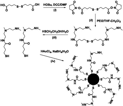
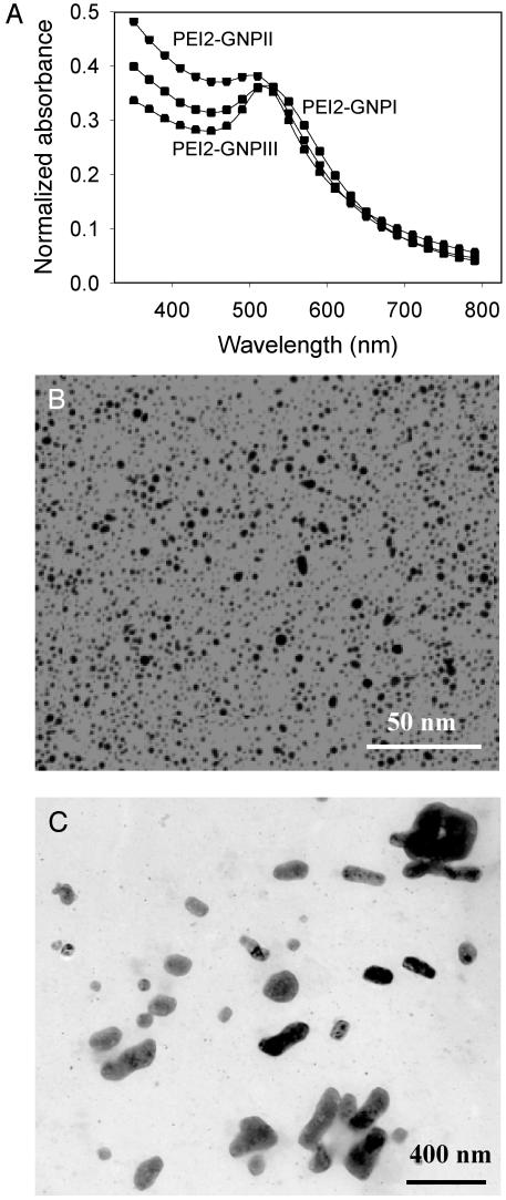
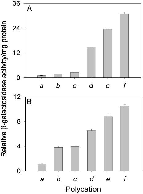
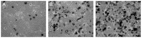
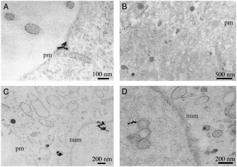
Branched polyethylenimine (PEI) chains with an average molecular mass of 2 kDa (PEI2) have been covalently attached to gold nanoparticles (GNPs), and the potency of the resulting PEI2-GNPs conjugates as vectors for the delivery of plasmid DNA into monkey kidney (COS-7) cells in the presence of serum in vitro has been systematically investigated. The transfection efficiencies vary as a function of the PEI/gold molar ratio in the conjugates, with the best one (PEI2-GNPII) being 12 times more potent than the unmodified polycation. This potency can be further doubled by adding amphiphilic N-dodecyl-PEI2 during complex formation with DNA. The resulting ternary complexes are at least 1 order of magnitude more efficient than the 25-kDa PEI, one of the premier polycationic gene-delivery vectors. Importantly, although unmodified PEI2 transfects just 4% of the cells, PEI2-GNPII transfects 25%, and the PEI2-GNPII/dodecyl-PEI2 ternary complex transfects 50% of the cells. The intracellular trafficking of the DNA complexes of these vectors, monitored by transmission electron microscopy, has detected the complexes in the nucleus <1 h after transfection.


Figures

Fig. 1.
Fig. 1.
Schematic representation of the synthetic route toward the conjugates of 2-kDa PEI and GNPs. Step i: conversion of 4,4′-dithiodibutyric acid to its bis-succinimide ester. HOSu, N-hydroxysuccinimide; DCC, dicyclohexylcarbodiimide; DMF, dimethylformamide. Step ii: reduction of the disulfides with 2-mercaptoethanol. Step iii: formation of PEI2–GNP conjugates by the reduction of hydrogen tetrachloroaurate in the presence of the thiol-modified PEI2. The last two reagents were mixed at three different molar ratios, 1:3, 3:1, and 10:1, to obtain the conjugates with different Au/PEI2 ratios (designated PEI2–GNPI, PEI2–GNPII, and PEI2–GNPIII, respectively). See Experimental Procedures for details.
Fig. 2.
Fig. 2.
Absorption spectra of the PEI2–GNP conjugates (A) and electron micrographs of PEI2–GNPII conjugates (B) and the polyplex of PEI2–GNPII with the plasmid DNA (gWiz Beta-Gal) formed at N/P = 150 (C). See Experimental Procedures for details.
Fig. 3.
Fig. 3.
(A) Extents of expression in COS-7 cell cultures of the β-gal gene mediated in the presence of serum by PEI2 (bars a–c) and PEI2–GNPII (bars d–f) at different N/P ratios. Bars a and d, N/P = 90; bars b and e, N/P = 120; bars c and f, N/P = 150. (B) Extents of expression in COS-7 cell cultures of the β-gal gene mediated in the presence of serum by PEI25 (bar a), dodecyl–PEI2 (bars b and c), PEI2–GNPII (bar d), and PEI2–GNPII + dodecyl–PEI2 (bars e and f). Bar a, N/P = 10; bar b, N/P = 20; bar c, N/P = 40; bar d, N/P = 150; bar e, N/P = 150 + 20; bar f, N/P = 150 + 40. Cells transfected with the plasmid in the absence of polycations showed no appreciable β-gal activity.
Fig. 4.
Fig. 4.
Histochemical staining with X-Gal of COS-7 cell cultures transfected in the presence of 10% serum with the β-gal gene mediated by PEI2 and its derivatives: PEI2 (N/P = 150) (A), PEI2–GNPII (N/P = 150) (B), and PEI2–GNPII + dodecyl–PEI2 (N/P = 150 + 40) (C).
Fig. 5.
Fig. 5.
Intracellular trafficking in COS-7 cell cultures of the polyplexes of PEI2–GNPII (A and B) and PEI2–GNPII + dodecyl–PEI2 (C and D) with plasmid DNA monitored by TEM 40 min after transfection. (A) Polyplexes are outside the cell and also being endocytosed. (B) Polyplexes have already been endocytosed. (C) Polyplexes entering the nucleus. (D) Polyplexes have entered the nucleus. Plasma membrane (pm) and nuclear membrane (num) are marked.

References

    1. Wang, L., Takabe, K., Bidlingmaier, S. M., Ill, C. R. & Verma, I. M. (1999) Proc. Natl. Acad. Sci. USA 96, 3906–3910. - PMC - PubMed
    1. Frater, A. J., Fidler, S. J. & McClure, M. O. (2002) Gene Ther. 9, 189–213.
    1. McNeish, I. & Seckl, M. J. (2002) Gene Ther. 9, 87–134.
    1. McCormick, F. (2001) Nat. Rev. Cancer 1, 130–140. - PubMed
    1. Marshall, E. (2000) Science 286, 2244–2245. - PubMed

Publication types