

Clusterin as a biomarker in murine and human intestinal neoplasia
- PMID: 12886021
- PMCID: PMC170952
- DOI: 10.1073/pnas.1233633100
Clusterin as a biomarker in murine and human intestinal neoplasia
Abstract
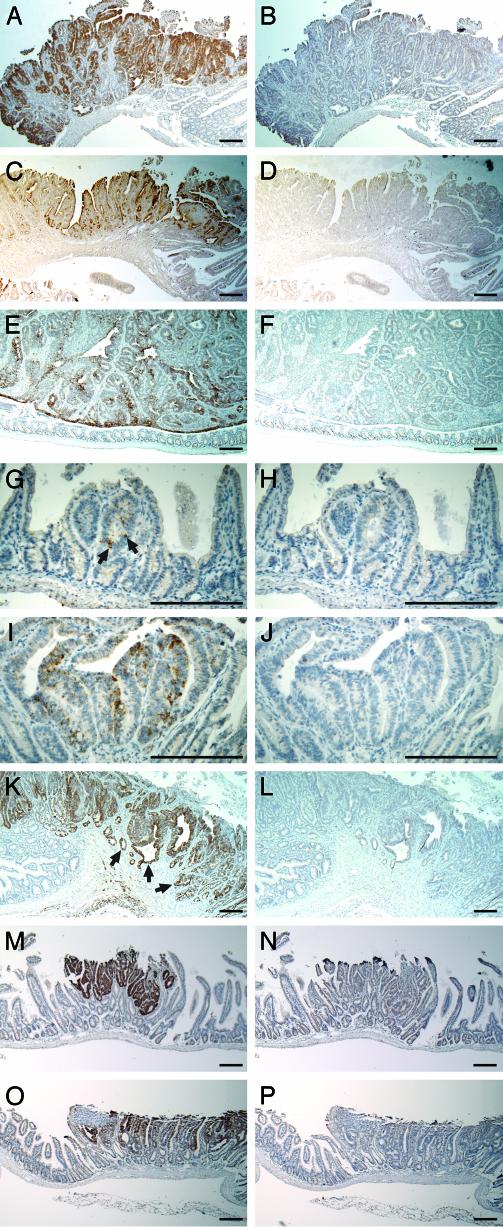
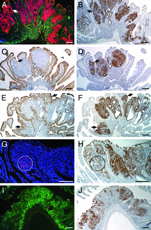
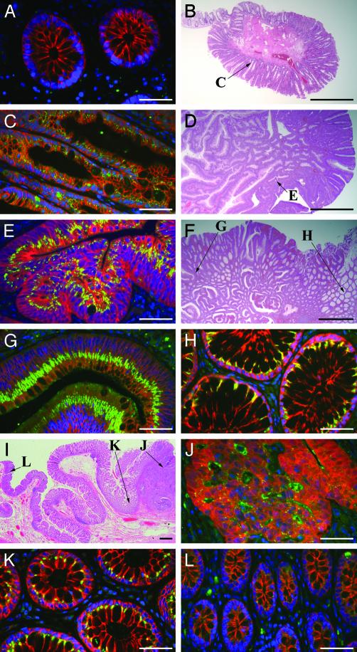
Early detection of colorectal cancer is critical for the management of this disease. Biomarkers for early detection of several cancers have been developed and applied clinically in recent years. We have sought to discover candidate biomarkers without the restricted choice of markers placed on microarrays, and without the biological complications of genetic and environmental heterogeneity. We have compared by cDNA subtraction two genetically matched sets of mice, one developing multiple intestinal neoplasia (C57BL/6J-ApcMin) and the other tumor-free (C57BL/6J). One prominent candidate biomarker, clusterin, was then subjected to a series of validation steps. In situ hybridization and immunohistochemistry were used to analyze clusterin expression at a cellular level on a series of murine intestinal and human colonic neoplasms. Elevated clusterin expression was characterized within certain regions of murine and human tumors regardless of tumor stage, location, or mode of initiation. The cells showing high clusterin levels generally lacked differentiation markers and adenomatous polyposis coli antigen. Tumor cells undergoing apoptosis expressed low levels of clusterin. Its specific expression patterns and correlation with cellular events during tumorigenesis make it a useful diagnostic tool in the mouse and a potential contributor to the set of biomarkers for early detection of human colon cancer.
Figures
Similar articles
-
Clusterin-mediated apoptosis is regulated by adenomatous polyposis coli and is p21 dependent but p53 independent.Cancer Res. 2004 Oct 15;64(20):7412-9. doi: 10.1158/0008-5472.CAN-04-2077. Cancer Res. 2004. PMID: 15492264
-
Clusterin expression in the early process of pancreas regeneration in the pancreatectomized rat.J Histochem Cytochem. 2003 Oct;51(10):1355-65. doi: 10.1177/002215540305101012. J Histochem Cytochem. 2003. PMID: 14500703
-
Dietary-induced ERbeta upregulation counteracts intestinal neoplasia development in intact male ApcMin/+ mice.Carcinogenesis. 2010 Feb;31(2):269-74. doi: 10.1093/carcin/bgp275. Epub 2009 Nov 27. Carcinogenesis. 2010. PMID: 19945967
-
Photoreceptor cells in the vitiligo mouse die by apoptosis. TRPM-2/clusterin expression is increased in the neural retina and in the retinal pigment epithelium.Invest Ophthalmol Vis Sci. 1995 Oct;36(11):2193-201. Invest Ophthalmol Vis Sci. 1995. PMID: 7558712
-
Clusterin (SGP-2, ApoJ) expression is downregulated in low- and high-grade human prostate cancer.Int J Cancer. 2004 Jan 1;108(1):23-30. doi: 10.1002/ijc.11496. Int J Cancer. 2004. PMID: 14618611
Cited by
-
Zinc Acts Synergistically with Berberine for Enhancing Its Efficacy as an Anti-cancer Agent by Inducing Clusterin-Dependent Apoptosis in HT-29 Colorectal Cancer Cells.Biol Trace Elem Res. 2023 Aug;201(8):3755-3773. doi: 10.1007/s12011-022-03460-8. Epub 2022 Nov 17. Biol Trace Elem Res. 2023. PMID: 36394793
-
17th International Mouse Genome Conference.Mamm Genome. 2004 Jul;15(7):509-14. doi: 10.1007/s00335-004-4001-9. Mamm Genome. 2004. PMID: 15366370 No abstract available.
-
Chromatin remodelling in damaged intestinal crypts orchestrates redundant TGFβ and Hippo signalling to drive regeneration.Nat Cell Biol. 2024 Dec;26(12):2084-2098. doi: 10.1038/s41556-024-01550-4. Epub 2024 Nov 15. Nat Cell Biol. 2024. PMID: 39548329
-
The Ins and Outs of Clusterin: Its Role in Cancer, Eye Diseases and Wound Healing.Int J Mol Sci. 2023 Aug 24;24(17):13182. doi: 10.3390/ijms241713182. Int J Mol Sci. 2023. PMID: 37685987 Free PMC article. Review.
-
The Influence of Clusterin Glycosylation Variability on Selected Pathophysiological Processes in the Human Body.Oxid Med Cell Longev. 2022 Aug 28;2022:7657876. doi: 10.1155/2022/7657876. eCollection 2022. Oxid Med Cell Longev. 2022. PMID: 36071866 Free PMC article. Review.
References
-
- American Cancer Society (2003) Cancer Facts & Figures 2003 (Am. Cancer Soc., Atlanta).
-
- Beart, R. W. (1995) in Clinical Oncology, eds. Abeloff, M. D., Armitage, J. O., Lichter, A. S. & Niederhuber, J. E. (Livingstone, New York), pp. 1267-1286.
-
- Neibergs, H. L., Hein, D. W. & Spratt, J. S. (2002) J. Surg. Oncol. 80, 204-213. - PubMed
-
- Saffran, D. C., Reiter, R. E., Jakobovits, A. & Witte, O. N. (1999) Cancer Metastasis Rev. 18, 437-449. - PubMed
-
- Burchardt, M., Burchardt, T., Shabsigh, A., de la Taille, A., Benson, M. C. & Sawczuk, I. (2000) Clin. Chem. 46, 595-605. - PubMed
Publication types
MeSH terms
Substances
Grants and funding
LinkOut - more resources
Full Text Sources
Other Literature Sources
Molecular Biology Databases
Research Materials
Miscellaneous
