

Induction of prolonged electrographic seizures in vitro has a defined threshold and is all or none: implications for diagnosis of status epilepticus
- PMID: 12887434
- PMCID: PMC2867609
- DOI: 10.1046/j.1528-1157.2003.51902.x
Induction of prolonged electrographic seizures in vitro has a defined threshold and is all or none: implications for diagnosis of status epilepticus
Abstract
Purpose: To study whether induction of prolonged (>30-min duration) in vitro electrographic seizure discharges resembling status epilepticus (SE) is graded or all-or-none, and to determine the critical factors mediating SE induction.
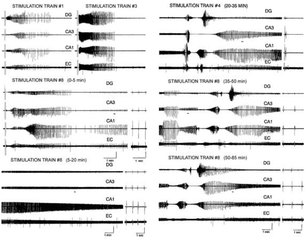
Methods: Prolonged electrographic seizure discharges were induced in combined hippocampal-entorhinal cortical (HEC) brain slices by electrical stimulation of the Schaeffer collaterals. Discharges were recorded by using field-potential electrodes in the dentate gyrus, CA3, CA1, and entorhinal cortex. Slices were prepared from rats that were (a). 21- to 30-day-old naive, (b). 60- to 120-day old naive, (c). epileptic, and (d). status post a prior traumatic brain injury.
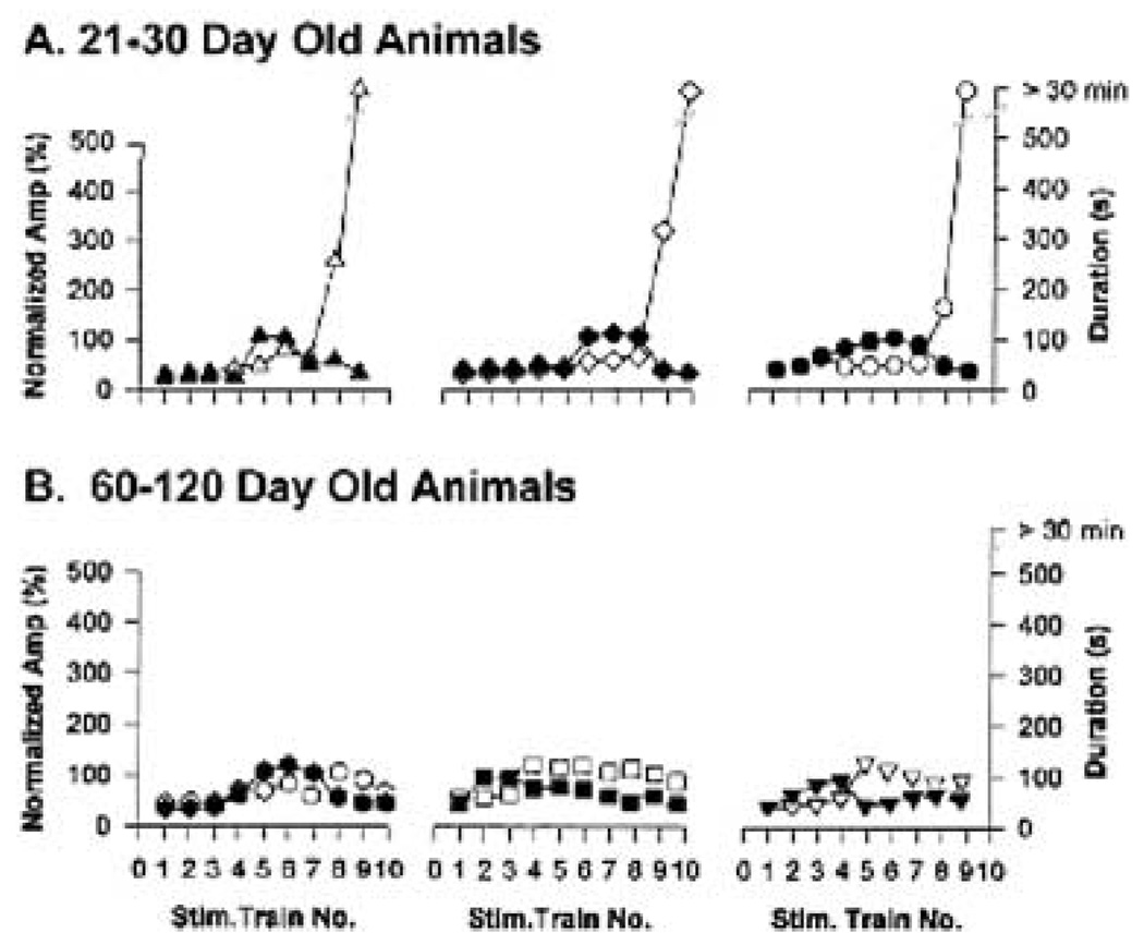
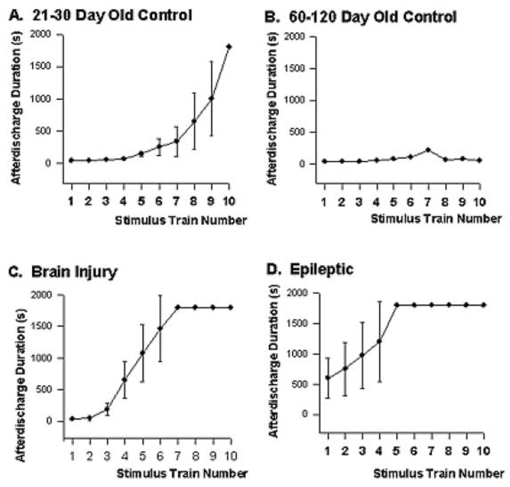
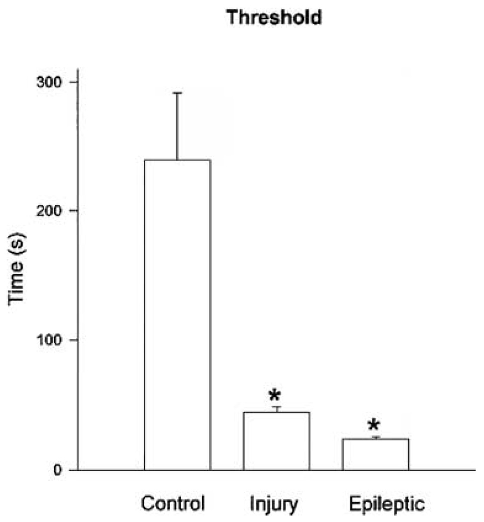
Results: Induction of SE discharges was dependent on the duration, but not amplitude of the preceding stimulus train-induced afterdischarge in HEC slices from 21- to 30-day-old control, brain-injured, and epileptic animals, but not from 60- to 120-day-old animals. In slices from 21- to 30-day-old control animals, once afterdischarges exceeded 4 min in duration, SE was induced in 50% of slices, and after >or=6 min 37 s seizure activity; SE was induced in 95% of slices. A defined SE threshold also was evident in brain-damaged rats, including rats in which an epileptic condition was induced by pilocarpine injection 4-16 weeks before recording, and rats subjected to a fluid percussive head trauma 1-8 weeks before recording. However, in these brain-damaged animals, mean SE threshold was considerably lower (24 and 44 s, respectively). HEC slices from 60- to 120-day-old controls for the brain-injured and epileptic animals did not develop SE even after 20 stimulations, demonstrating the pronounced effect of brain injury and epilepsy on the development of SE in the HEC slice preparation compared with that in age-matched controls.
Conclusions: In vitro, SE discharges have a defined temporal threshold for initiation. Once a seizure exceeds 6-7 min in duration in control animals, and 30-55 s in brain-damaged animals, the probability of SE induction is greatly increased. This demonstrates that brain injury lowers the afterdischarge duration required to produce SE and suggests that brains injured from trauma or SE are more susceptible to develop status epilepticus.
Figures
Similar articles
-
Long-duration self-sustained epileptiform activity in the hippocampal-parahippocampal slice: a model of status epilepticus.J Neurophysiol. 1995 Nov;74(5):2028-42. doi: 10.1152/jn.1995.74.5.2028. J Neurophysiol. 1995. PMID: 8592194
-
Generation and propagation of epileptiform discharges in a combined entorhinal cortex/hippocampal slice.J Neurophysiol. 1993 Nov;70(5):1962-74. doi: 10.1152/jn.1993.70.5.1962. J Neurophysiol. 1993. PMID: 8294965
-
Responses of deep entorhinal cortex are epileptiform in an electrogenic rat model of chronic temporal lobe epilepsy.J Neurophysiol. 1998 Jul;80(1):230-40. doi: 10.1152/jn.1998.80.1.230. J Neurophysiol. 1998. PMID: 9658044
-
Network and pharmacological mechanisms leading to epileptiform synchronization in the limbic system in vitro.Prog Neurobiol. 2002 Oct;68(3):167-207. doi: 10.1016/s0301-0082(02)00077-1. Prog Neurobiol. 2002. PMID: 12450487 Review.
-
Do interictal discharges promote or control seizures? Experimental evidence from an in vitro model of epileptiform discharge.Epilepsia. 2001;42 Suppl 3:2-4. doi: 10.1046/j.1528-1157.2001.042suppl.3002.x. Epilepsia. 2001. PMID: 11520313 Review.
Cited by
-
Generalised convulsive status epilepticus: an overview.Postgrad Med J. 2006 Nov;82(973):723-32. doi: 10.1136/pgmj.2005.043182. Postgrad Med J. 2006. PMID: 17099091 Free PMC article. Review.
-
Xenon LFP Analysis Platform Is a Novel Graphical User Interface for Analysis of Local Field Potential From Large-Scale MEA Recordings.Front Neurosci. 2022 Jul 1;16:904931. doi: 10.3389/fnins.2022.904931. eCollection 2022. Front Neurosci. 2022. PMID: 35844228 Free PMC article.
References
-
- DeLorenzo RJ, Towne AR, Pellock JM, et al. Status epilepticus in children, adults, and the elderly. Epilepsia. 1992;33:14–24. - PubMed
-
- Hauser WA. Status epilepticus: epidemiological considerations. Neurology. 1990;40 suppl 2:9–13. - PubMed
-
- Smith DH, Lowenstein DH, Gennerelli TA, et al. Persistent memory dysfunction is associated with bilateral hippocampal damage following experimental brain injury. Neurosci Lett. 1994;168:151–154. - PubMed
-
- Traub RD, Miles R, Wong RKS. Models of synchronized hippocampal bursts in the presence of inhibition, I: single population events. J Neurophysiol. 1987;58:739–751. - PubMed
-
- Epilepsy Foundation of America Working Group on Status Epilepticus. Treatment of convulsive status epilepticus. JAMA. 1993;270:854–859. - PubMed
Publication types
MeSH terms
Grants and funding
LinkOut - more resources
Full Text Sources
Miscellaneous
