. 2003 Aug 1;31(15):4285-92.
doi: 10.1093/nar/gkg655.

Mechanisms of P/CAF auto-acetylation

Affiliations

Mechanisms of P/CAF auto-acetylation

Helena Santos-Rosa et al. Nucleic Acids Res. .

Abstract

P/CAF is a histone acetyltransferase enzyme which was originally identified as a CBP/p300-binding protein. In this manuscript we report that human P/CAF is acetylated in vivo. We find that P/CAF is acetylated by itself and by p300 but not by CBP. P/CAF acetylation can be an intra- or intermolecular event. The intermolecular acetylation requires the N-terminal domain of P/CAF. The intramolecular acetylation targets five lysines (416-442) at the P/CAF C-terminus, which are in the nuclear localisation signal (NLS). Finally, we show that acetylation of P/CAF leads to an increment of its histone acetyltransferase (HAT) activity. These findings identify a new post-translation modification on P/CAF which may regulate its function.


Figures

Figure 1
Figure 1
P/CAF is an acetyl-protein in vivo. (A) The antibody against Acetyl-Lys recognises acetylated P/CAF; 0.5 µg of recombinant GST-P/CAF was incubated for 0–2 h with acetyl-CoA, and the acetylated products were analysed by western blotting using an antibody that recognises acetylated lysines (anti-acetyl-Lys). The bottom band in the gel (indicated by a small arrow) corresponds to a GST-P/CAF degradation product. (B) P/CAF is acetylated in vivo. U2Os cells were transfected with 10 µg of Pcx-P/CAF-Flag, Pcx-P/CAFΔHAT-Flag and pCDNA3-HDAC1-Flag plasmids. Whole cell extracts from transfected cells were prepared, and Flag-proteins from them were immunoprecipitated using an anti-Flag antibody. The immunocomplexes were tested for P/CAF acetylation status by western blotting, using the antibody, which recognises acetylated lysines (anti-acetyl- Lys) (bottom part), and an antibody against the Flag-tag (top part). (C) Endogenous P/CAF is acetylated in vivo. P/CAF from C2C12 cells was immunoprecipitated using an anti P/CAF antibody or the preimmune serum (as a control), and the immunocomplexes were analysed for their acetylation level by western blotting, using the anti-acetyl-Lys antibody.
Figure 2
Figure 2
P/CAF is acetylated in cis and in trans in vivo. (A) P/CAF is acetylated in trans. U2Os cells were transfected with 10 µg of pCX-GFP-P/CAF-Flag and pCX-P/CAFΔHAT-Flag plasmids. Whole cell extracts from transfected cells were prepared and Flag-proteins from them were immunoprecipitated using an anti-Flag antibody. The immunocomplexes were tested for P/CAF acetylation status by western blotting, using the anti-acetyl-Lys antibody (bottom part of the figure) and an antibody against the Flag tag (top part). Positions of the acetylated GFP-P/CAF-Flag and acetylated P/CAFΔHAT-Flag are indicated by arrows. (B) P/CAF N-terminal domain is required for P/CAF trans acetylation in vivo. U2OS cells were transfected with 10 µg of pCX-P/CAF(FL)-Flag and pCX-P/CAFΔHAT (352–832)-Flag plasmids. Whole cell extracts were prepared, Flag proteins were immunoprecipitated by using the Flag antibody and P/CAF acetylation was analysed by western blotting using the anti-acetyl-Lys antibody (bottom part) and the anti-Flag antibody as a control (top part). The small arrow indicates where acetylated P/CAFΔHAT (352–832)-Flag should be found. (C) Deletion of the N-terminal domain does not affect P/CAF-P/CAF interaction. GST, GST-P/CAF and GST-P/CAF (352–832) fusion proteins were expressed and the binding to P/CAF FL in vitro translated and radiolabelled was determined by an in vitro pull down experiment (see Materials and Methods). Input represents 25% of total P/CAF input. The arrow indicates the P/CAF position in the gel.
Figure 3
Figure 3
P/CAF is acetylated in cis at the C-terminus. (A) Recombinant GST-P/CAF(FL), GST-P/CAF (352–832) and GST-P/CAFΔHAT (352–832) were expressed in E.coli and purified. Fusion proteins were used in an in vitro acetylation assay. Acetylated proteins were visualised by fluorography. (B) HAT assay of GST-P/CAF(FL) and GST-P/CAF (352–832) recombinant proteins. (C) U2Os cells were transfected with 10 µg of pCX-P/CAF (352–832)-Flag and pCX-P/CAFΔHAT (352–832)-Flag plasmids. Whole cell extracts were prepared, Flag proteins were immunoprecipitated by using the Flag antibody and the immunocomplexes were tested for P/CAF acetylation status by western blotting, using the antibody which recognises acetylated lysines (anti-acetyl-Lys) (bottom part) and an antibody against the Flag-tag (top part). The position of the inactive protein is indicated by an arrow.
Figure 4
Figure 4
P/CAF is acetylated by p300 in vivo. (A) PCX-P/CAF-Flag, PCX-P/CAFΔHAT-Flag and pCDNA3-p300-HA plasmids were transfected into U2OS cells. Proteins were immunoprecipitated using antibodies against HA (lanes 1, 4 and 7) and Flag (lanes 2, 3, 5 and 6) tags. The acetylation level of P/CAF was analysed by western blotting using the anti-acetyl-Lys antibody (bottom panel). (B) PCX-P/CAFΔHAT-Flag, pCDNA3-p300-HA and pCDNA3-CBP-HA plasmids were transfected into U2OS cells. The proteins were immunoprecipitated using antibodies against HA and Flag tags and the acetylated status of P/CAF was analysed by western blotting using the anti-acetyl-Lys antibody. (C) IP-HAT assay. U2OS cells were transfected with pCDNA3-p300-HA, pCDNA3-CBP-HA or the empty vector. CBP and p300 were immunoprecipitated using antibodies against HA and the HAT activity associated with the immunocomplexes was determined by liquid HAT assay.
Figure 4
Figure 4
P/CAF is acetylated by p300 in vivo. (A) PCX-P/CAF-Flag, PCX-P/CAFΔHAT-Flag and pCDNA3-p300-HA plasmids were transfected into U2OS cells. Proteins were immunoprecipitated using antibodies against HA (lanes 1, 4 and 7) and Flag (lanes 2, 3, 5 and 6) tags. The acetylation level of P/CAF was analysed by western blotting using the anti-acetyl-Lys antibody (bottom panel). (B) PCX-P/CAFΔHAT-Flag, pCDNA3-p300-HA and pCDNA3-CBP-HA plasmids were transfected into U2OS cells. The proteins were immunoprecipitated using antibodies against HA and Flag tags and the acetylated status of P/CAF was analysed by western blotting using the anti-acetyl-Lys antibody. (C) IP-HAT assay. U2OS cells were transfected with pCDNA3-p300-HA, pCDNA3-CBP-HA or the empty vector. CBP and p300 were immunoprecipitated using antibodies against HA and the HAT activity associated with the immunocomplexes was determined by liquid HAT assay.
Figure 5
Figure 5
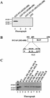
Mapping of P/CAF cis-acetylation sites. (A) Recombinant GST-P/CAF(352–658) (lane 1), GST-P/CAF(489–658) (lane 2), GST-P/CAFΔHAT(352–658) (lane 3) and GST (lane 4) were tested for autoacetylation in an in vitro acetylation assay. The acetylated proteins were visualised by fluorography. (B) Schematic representation of the P/CAF (352–658) region (acetylated lysines are indicated). (C) Fluorograph showing P/CAF acetylation of the GST-P/CAF (352–658) wt (lane 2) and punctual mutants (lanes 3–9) indicated on the top of the figure. Lane 1 corresponds to the protein molecular weight marker.
Figure 6
Figure 6
The acetylated lysines in cis are essential for the P/CAF NLS function. C2C12 cells were transfected with pCX-GFP-P/CAF(FL) and pCX-GFP-P/CAF(FL)(Ks-Rs) in which lysines 428, 430, 441 and 442 have been mutated to arginines. Localisation of the expressed proteins was detected before and after differentiation of C2C12 cells by fluorescence.
Figure 7
Figure 7
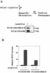
P/CAF auto-acetylation and HAT activity. (A) GST-P/CAF (352–658) or the non-acetylable P/CAF mutant [GST-P/CAF (352–658)K-R mutated at 416, 428, 430, 441 and 442 lysines] were incubated in the presence or absence of acetyl-CoA for 5 min, then purified H3 was added to the reaction and the P/CAF HAT activity on H3 was analysed by fluorography. (B) The results in (A) were quantified using a Molecular Dynamics machine and normalised relative to the values obtained in absence of acetyl-CoA (value 1).

Similar articles

Cited by

References

    1. Konberg R.D. and Lorch,Y. (1999) Chromatin-modifying and remodelling complexes. Curr. Opin. Genet. Dev., 9, 148–151. - PubMed
    1. Kingston R.E. and Narlikar,G.J. (1999) ATP-dependent remodelling and acetylation as regulators of chromatin fluidity. Genes Dev., 13, 2339–2352. - PubMed
    1. Wu J. and Grunstein,M. (2000) 25 years after the nucleosome model: chromatin modifications. Trends Biochem. Sci., 25, 619–623. - PubMed
    1. Berger S. (2002) Histone modifications in transcriptional regulation. Curr. Opin. Genet. Dev., 12, 142–148. - PubMed
    1. Hebbes T.R., Thorne,A.W. and Crane-Robinson,C. (1988) A direct link between core histone acetylation and transcriptionally active chromatin. EMBO J., 7, 1395–1402. - PMC - PubMed

Publication types

MeSH terms