. 2003 Aug 1;31(15):4293-303.
doi: 10.1093/nar/gkg650.

Functional characterization of cis- and trans-regulatory elements involved in expression of phospholipid hydroperoxide glutathione peroxidase

Affiliations

Functional characterization of cis- and trans-regulatory elements involved in expression of phospholipid hydroperoxide glutathione peroxidase

Christoph Ufer et al. Nucleic Acids Res. .

Abstract

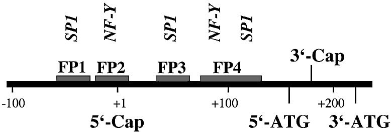
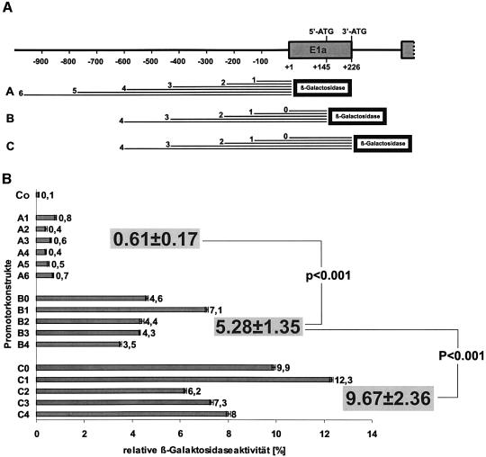
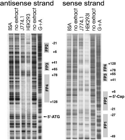
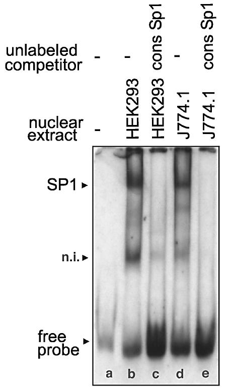
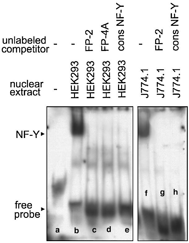
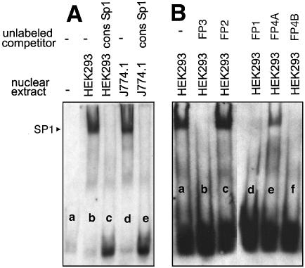
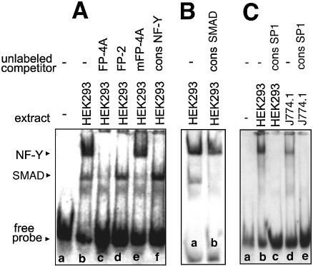
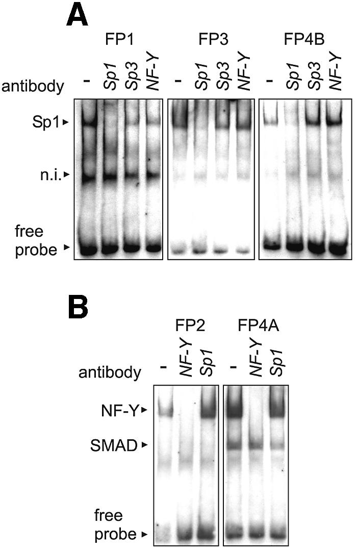
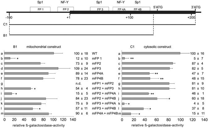
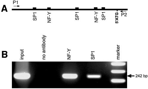
Phospholipid hydroperoxide glutathione peroxidase (phGPx) is a member of the seleno glutathione peroxidase family that is comprised of five selenoproteins capable of reducing hydroperoxy lipids to the corresponding alcohols. The enzyme has been implicated in antioxidative defense, but its high expression level in testicular tissue suggests a more specific function during sperm maturation. The phGPx is encoded for by a joint sperm nucleus/phGPx gene (sn/phGPx) and can be expressed as a mitochondrial or cytosolic isoform. Although sn/phGPx genes have been cloned from various mammalian species expression regulation of the enzyme has not been studied in detail. We investigated the 5'-flanking region of the murine sn/phGPx gene and observed basic promoter activity in a 200 bp region localized immediately upstream of the translational initiation site of the cytosolic isoform (3'-ATG). DNase protection assays indicated the presence of five distinct protein-binding regions and electrophoretic mobility shift assays and supershift experiments revealed binding of stimulating protein 1 (SP1), nuclear factor Y (NF-Y) and members of the SMAD family. Site-directed mutagenesis of the consensus binding sequences abolished in vitro transcription factor binding. Expression of reporter genes was most effectively impaired when SP1/SP3 and NF-Y binding site-deficient constructs were tested. Chromatin immunoprecipitation suggested the in vivo relevance of these transcription factors. Our data indicate that the basic phGPx promoter constitutes a 200 bp oligonucleotide, which is localized immediately upstream of the 3'-ATG and involves functional SP1/SP3, NF-Y and SMAD binding sites. The corresponding trans-regulatory proteins may contribute to differential expression regulation of the mitochondrial and cytosolic phGPx isoforms.


Figures

None
Scheme 1. Multiple transcription initiation sites in mouse and rat tissues. The location of the murine translational start codons at +145 (5′-ATG) and ATG at +226 (3′-ATG) are shown. Thick arrows indicate different transcription initiation sites identified in mice for the mitochondrial and cytosolic phGPx isoform. The most 5′ transcription start site identified so far in mice (testis) was set +1 (33). The asterisk indicates two additional transcription start sites reported for various murine organs (15). The two windows of transcription start sites identified in rat tissues (11) are indicated by brackets.
None
Scheme 2. Functional cis-acting elements involved in transcriptional regulation of mitochondrial and cytosolic phGPx isoforms. The 5′-ATG represents the translational initiation site for the mitochondrial isoform whereas translation of the cytosolic enzyme starts at the 3′-ATG. The transcriptional initiation sites for the long (mitochondrial isoform) and the short messenger (cytosolic messenger) are indicated by the 5′- and the 3′-Cap, respectively.
Figure 1
Figure 1
Functional promoter studies of the 5′-flanking region of the joint ph/snGPx gene. Different sections of the 5′-flanking region of the joint ph/snGPx gene were ligated into a β-galactosidase-based reporter gene and HEK293 cells were transfected with the different constructs using the calcium phosphate transfection method. In order to correct the β-galactosidase activity measured for transfection efficiency, cotransfections with luciferase containing control plasmid (pGL4) were carried out. (A) Length (bp) of the PCR fragments of the 5′-flanking regions used for reporter gene construction. (B) Relative β-galactosidase activity of the different promoter constructs corrected for luciferase activity. Co-negative control (pBlueTopo without insert).
Figure 2
Figure 2
DNA footprinting of the 5′-flanking region of the joint ph/snGPx gene. DNase protection assays (footprints) were carried out as described in Materials and Methods. A genomic fragment containing the 5′-UTR of the cytosolic phGPx isoform as well as ∼100 bp of the 5′-flanking region of the phGPx gene (–99 to +228 bp) was used. The areas of quenched signals indicate the protein-binding sites (FP-1 to FP-4). Unspecific quenching was tested using BSA but we did not observe any unspecific effects. The sequences of the footprints were determined with the G+A ladder.
Figure 3
Figure 3
EMSAs using an oligonucletide of FP-1 as labeled probe. The labeled oligonucleotide dimer (GTA AGC CCA GCC CCG CCC AAG CCG TCC CTT CAT TCA) containing the binding sequence for SP1 was incubated with nuclear extracts of HEK293 cells (10 µg of protein) or J774.1 cells (10 µg of protein) in the presence or absence of unlabeled SP1 consensus oligonucleotides (9.6 pmol) as described in Materials and Methods. After 30 min of incubation at 4°C the entire binding sample was applied to polyacrylamide gel electrophoresis. The arrows indicate the shift bands. n.i., not identified.
Figure 4
Figure 4
EMSAs using an oligonucletide of FP-2 as labeled probe. The labeled oligonucleotide dimer (ATT CAG GCT TCC CAT TGG CTG CAG GGG CCT CGC GTC) containing the binding sequence for NF-Y was incubated with nuclear extracts of HEK293 cells (10 µg of protein) or J774.1 cells (10 µg of protein) in the presence or absence of unlabeled competitors or NF-Y consensus oligonucleotides (9.6 pmol) as described in Materials and Methods. After 30 min of incubation at 4°C the entire binding sample was applied to polyacrylamide gel electrophoresis. The arrows indicate the shift bands.
Figure 5
Figure 5
EMSAs using an oligonucletide of FP-3 as labeled probe. The labeled oligonucleotide dimer (AAT AAG AGA CGT CAG TGG GCG TGC CCG AGG GCG GGC) containing the binding sequence for SP1 was incubated with nuclear extracts of HEK293 cells (10 µg of protein) or J774.1 cells (10 µg of protein) in the presence or absence of unlabeled (A) SP1 consensus oligonucleotides or (B) competitors (9.6 pmol) as described in Materials and Methods. After 30 min of incubation at 4°C the entire binding sample was applied to polyacrylamide gel electrophoresis. The arrow indicates the shift bands.
Figure 6
Figure 6
EMSAs using an oligonucletide of FP-4 as labeled probe. The labeled oligonucleotide dimer (CCA TAC TCG GCC TCG CGC GTC CAT TGG TCG GCT GCG) containing the overlapping binding sequences for SMAD and NF-Y (A and B) or the labeled oligonucleotide (TCG GCT GCG TGA GGG GAG GAG CCG CTG GCT CCG GCC) containing the binding sequence for SP1 (C) was incubated with nuclear extracts of HEK293 cells (10 µg of protein) or J774.1 cells (10 µg of protein) in the presence or absence of unlabeled competitors or consensus oligonucleotides (9.6 pmol) as described in Materials and Methods. After 30 min of incubation at 4°C the entire binding sample was applied to polyacrylamide gel electrophoresis. The arrows indicate the shift bands.
Figure 7
Figure 7
Supershift assays using antibodies against SP1, SP3 and NF-Y. EMSA experiments were carried out as described in the captions to Figures 3–6. For the supershift experiments shown in this figure, specific antibodies raised against SP1, SP3 and NF-Y were added to the incubation mixtures. (A) Footprints FP-1, FP-3 and FP-4B containing SP1/SP3 binding sites were analyzed. (B) Footprints FP-2 and FP-4A containing NF-Y binding sites were analyzed. n.i., not identified.
Figure 8
Figure 8
Functional promoter studies using mutated reporter gene constructs. Site-directed mutagenesis of the phGPx promoter was performed as described in Materials and Methods. A mixture of each of 1 µg of β-galactosidase promoter construct and 1 µg of pGL3 luciferase plasmid were transfected into HEK293 cells with FuGene 6 (see Materials and Methods). The β-galactosidase activity was normalized for the luciferase activity. Values are means ± SEM for three determinations given as a percentage compared with the wild-type construct. (Top) Localization of footprints (FP-1 to FP-4) and functional transcription factor binding sites in the 5′-flanking region of the sn/phGPx gene and the length of the reporter gene constructs B1 and C1. The major transcription initiation site for the mitochondrial phGPx isoform was set +1. Rectangular arrows indicate heterogeneity of transcription initiation sites. 5′-ATG (translational initiation site for the mitochondrial phGPx) and 3′-ATG (translational initiation site for the cytosolic phGPx) are also indicated. (Bottom) Relative promoter activity of wild-type (WT) and mutant reporter gene constructs. *P < 0.05; **P < 0.01 compared with the wild-type construct.
Figure 9
Figure 9
Chromatin immunoprecipitation of relevant transcription factors. Chromatin immunoprecipitation was carried out as described in Materials and Methods. (A) Localization of the relevant transcription factor binding sites in the promoter of the ph/snGPx gene. The arrows indicate the positions of the primers used for amplification of the immunoprecipitated DNA. (B) Sheared DNA/protein complexes were precipitated with antibodies against NF-Y and SP1 and precipitated genomic DNA was amplified. Input, sheared DNA prior to immunoprecipitation. A control immunoprecipitation using an irrelevant antibody (anti-15-LOX antibody) did not show any PCR signal (see Supplementary Material).

Similar articles

Cited by

References

    1. Flohé L. (1989) The selenoprotein glutathione peroxidase. In Dolphin,D., Poulson,R. and Avramovic,O. (eds), Glutathione: Chemical, Biochemical and Medical Aspects—Part A. John Wiley and Sons, New York, pp. 643–731.
    1. Arthur J.R. (2000) The glutathione peroxidases. Cell. Mol. Life Sci., 57, 1825–1835. - PMC - PubMed
    1. Behne D. and KyriakopoulosA. (2001) Mammalian selenium-containing proteins. Annu. Rev. Nutr., 21, 453–473. - PubMed
    1. Kuhn H. and Borchert,A. (2002) Regulation of enzymatic lipid peroxidation. The interplay of peroxidizing and peroxide reducing enzymes. Free Radic. Biol. Med., 33, 154–172. - PubMed
    1. Ursini F., Maiorino,M., Valente,M., Ferri,L. and Gregolin,C. (1982) Purification from pig liver of a protein which protects liposomes and biomembranes from peroxidative degradation and exhibits glutathione peroxidase activity on phosphatidylcholine hydroperoxides. Biochim. Biophys. Acta, 710, 197–211. - PubMed

Substances