

Hydropathic analysis of the free energy differences in anthracycline antibiotic binding to DNA
- PMID: 12888500
- PMCID: PMC169931
- DOI: 10.1093/nar/gkg645
Hydropathic analysis of the free energy differences in anthracycline antibiotic binding to DNA
Abstract
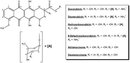
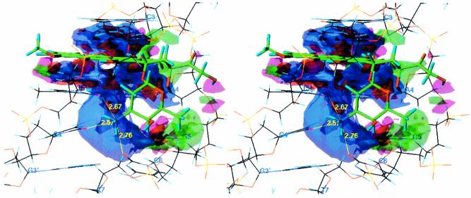
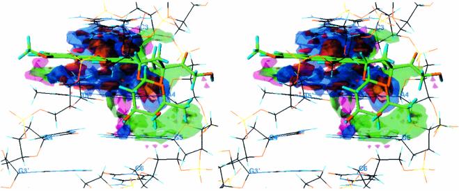
Molecular models of six anthracycline antibiotics and their complexes with 32 distinct DNA octamer sequences were created and analyzed using HINT (Hydropathic INTeractions) to describe binding. The averaged binding scores were then used to calculate the free energies of binding for comparison with experimentally determined values. In parsing our results based on specific functional groups of doxorubicin, our calculations predict a free energy contribution of -3.6 +/- 1.1 kcal x mol(-1) (experimental -2.5 +/- 0.5 kcal x mol(-1)) from the groove binding daunosamine sugar. The net energetic contribution of removing the hydroxyl at position C9 is -0.7 +/- 0.7 kcal x mol(-1) (-1.1 +/- 0.5 kcal x mol(-1)). The energetic contribution of the 3' amino group in the daunosamine sugar (when replaced with a hydroxyl group) is -3.7 +/- 1.1 kcal x mol(-1) (-0.7 +/- 0.5 kcal x mol(-1)). We propose that this large discrepancy may be due to uncertainty in the exact protonation state of the amine. The energetic contribution of the hydroxyl group at C14 is +0.4 +/- 0.6 kcal x mol(-1) (-0.9 +/- 0.5 kcal x mol(-1)), largely due to unfavorable hydrophobic interactions between the hydroxyl oxygen and the methylene groups of the phosphate backbone of the DNA. Also, there appears to be considerable conformational uncertainty in this region. This computational procedure calibrates our methodology for future analyses where experimental data are unavailable.
Figures
Similar articles
-
Parsing the free energy of anthracycline antibiotic binding to DNA.Biochemistry. 1996 Feb 20;35(7):2047-53. doi: 10.1021/bi952812r. Biochemistry. 1996. PMID: 8652545
-
A computational model for anthracycline binding to DNA: tuning groove-binding intercalators for specific sequences.J Med Chem. 2004 Mar 11;47(6):1360-74. doi: 10.1021/jm030529h. J Med Chem. 2004. PMID: 14998326
-
Electrostatic and non-electrostatic contributions to the binding free energies of anthracycline antibiotics to DNA.J Mol Biol. 1997 Nov 28;274(2):253-67. doi: 10.1006/jmbi.1997.1399. J Mol Biol. 1997. PMID: 9398531
-
Comparison of DNA sequence selectivity of anthracycline antibiotics and their 3'-hydroxylated analogs.Chem Biol Interact. 1996 Mar 25;100(2):165-76. doi: 10.1016/0009-2797(96)03697-6. Chem Biol Interact. 1996. PMID: 8646789
-
Review on the binding of anticancer drug doxorubicin with DNA and tRNA: Structural models and antitumor activity.J Photochem Photobiol B. 2016 May;158:274-9. doi: 10.1016/j.jphotobiol.2016.02.032. Epub 2016 Mar 4. J Photochem Photobiol B. 2016. PMID: 26971631 Review.
Cited by
-
HINT, a code for understanding the interaction between biomolecules: a tribute to Donald J. Abraham.Front Mol Biosci. 2023 Jun 7;10:1194962. doi: 10.3389/fmolb.2023.1194962. eCollection 2023. Front Mol Biosci. 2023. PMID: 37351551 Free PMC article. Review.
-
Cooperative effects on the formation of intercalation sites.Nucleic Acids Res. 2004 Sep 1;32(15):4696-703. doi: 10.1093/nar/gkh788. Print 2004. Nucleic Acids Res. 2004. PMID: 15342790 Free PMC article.
-
Autoregulation of antibiotic biosynthesis by binding of the end product to an atypical response regulator.Proc Natl Acad Sci U S A. 2009 May 26;106(21):8617-22. doi: 10.1073/pnas.0900592106. Epub 2009 May 7. Proc Natl Acad Sci U S A. 2009. PMID: 19423672 Free PMC article.
-
Daunorubicin-Loaded DNA Origami Nanostructures Circumvent Drug-Resistance Mechanisms in a Leukemia Model.Small. 2016 Jan 20;12(3):308-20. doi: 10.1002/smll.201502118. Epub 2015 Nov 19. Small. 2016. PMID: 26583570 Free PMC article.
-
A new bisintercalating anthracycline with picomolar DNA binding affinity.J Med Chem. 2005 Dec 29;48(26):8209-19. doi: 10.1021/jm050902g. J Med Chem. 2005. PMID: 16366602 Free PMC article.
References
-
- Weiss R.B. (1992) The anthracyclines: will we ever find a better doxorubicin? Semin. Oncol., 19, 670–686. - PubMed
-
- Momparler R.L., Karon,M., Siegel,S.E. and Avila,F. (1976) Effect of adriamycin on DNA, RNA and protein synthesis in cell-free systems and intact cells. Cancer Res., 36, 2891–2895. - PubMed
-
- Tewey K.M., Rowe,T.C., Yang,L., Halligan,B.D. and Lui,L.F. (1984) Adriamycin-induced DNA damage mediated by mammalian DNA topoisomerase II. Science, 226, 466–468. - PubMed
-
- Schneider E., Hsiang,Y. and Lui,L.F. (1990) DNA topoisomerases as anticancer drug targets. Adv. Pharmacol., 21, 149–183. - PubMed
-
- Sander M. and Tsieh,T.-S. (1983) Double strand DNA cleavage by type II DNA topoisomerase from Drosophila melanogaster. J. Biol. Chem., 258, 8421–8428. - PubMed
Publication types
MeSH terms
Substances
LinkOut - more resources
Full Text Sources
Research Materials
Miscellaneous
