

Global methylation screening in the Arabidopsis thaliana and Mus musculus genome: applications of virtual image restriction landmark genomic scanning (Vi-RLGS)
- PMID: 12888509
- PMCID: PMC169878
- DOI: 10.1093/nar/gkg488
Global methylation screening in the Arabidopsis thaliana and Mus musculus genome: applications of virtual image restriction landmark genomic scanning (Vi-RLGS)
Abstract
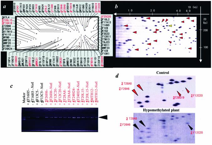
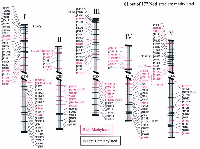
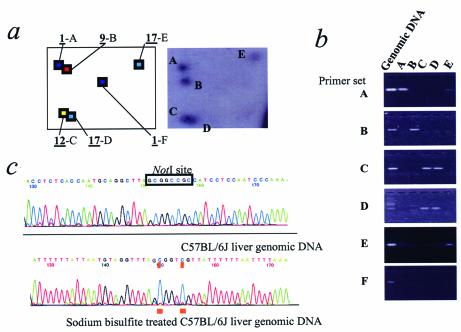
Understanding the role of 'epigenetic' changes such as DNA methylation and chromatin remodeling has now become critical in understanding many biological processes. In order to delineate the global methylation pattern in a given genomic DNA, computer software has been developed to create a virtual image of restriction landmark genomic scanning (Vi-RLGS). When using a methylation- sensitive enzyme such as NotI as the restriction landmark, the comparison between real and in silico RLGS profiles of the genome provides a methylation map of genomic NotI sites. A methylation map of the Arabidopsis genome was created that could be confirmed by a methylation-sensitive PCR assay. The method has also been applied to the mouse genome. Although a complete methylation map has not been completed, a region of methylation difference between two tissues has been tested and confirmed by bisulfite sequencing. Vi-RLGS in conjunction with real RLGS will make it possible to develop a more complete map of genomic sites that are methylated or demethylated as a consequence of normal or abnormal development.
Figures


Similar articles
-
Restriction landmark genomic scanning (RLGS) spot identification by second generation virtual RLGS in multiple genomes with multiple enzyme combinations.BMC Genomics. 2007 Nov 30;8:446. doi: 10.1186/1471-2164-8-446. BMC Genomics. 2007. PMID: 18053125 Free PMC article.
-
Restriction landmark genome scanning.Methods Mol Biol. 2011;791:101-12. doi: 10.1007/978-1-61779-316-5_8. Methods Mol Biol. 2011. PMID: 21913074
-
Restriction landmark genomic scanning for DNA methylation in cancer: past, present, and future applications.Anal Biochem. 2002 Aug 15;307(2):191-201. doi: 10.1016/s0003-2697(02)00033-7. Anal Biochem. 2002. PMID: 12202234 Review.
-
Restriction landmark genome scanning method using isoschizomers (MspI/HpaII) for DNA methylation analysis.Electrophoresis. 2006 Jul;27(14):2846-56. doi: 10.1002/elps.200500776. Electrophoresis. 2006. PMID: 16637018
-
Epigenetics: application of virtual image restriction landmark genomic scanning (Vi-RLGS).FEBS J. 2008 Apr;275(8):1608-16. doi: 10.1111/j.1742-4658.2008.06329.x. Epub 2008 Mar 7. FEBS J. 2008. PMID: 18331348 Review.
Cited by
-
Aberrant methylation of the TDMR of the GTF2A1L promoter does not affect fertilisation rates via TESE in patients with hypospermatogenesis.Asian J Androl. 2013 Sep;15(5):634-9. doi: 10.1038/aja.2013.56. Epub 2013 Jun 17. Asian J Androl. 2013. PMID: 23770943 Free PMC article.
-
Tissue specific differentially methylated regions (TDMR): Changes in DNA methylation during development.Genomics. 2009 Feb;93(2):130-9. doi: 10.1016/j.ygeno.2008.09.003. Epub 2008 Nov 13. Genomics. 2009. PMID: 18952162 Free PMC article.
-
Comparative isoschizomer profiling of cytosine methylation: the HELP assay.Genome Res. 2006 Aug;16(8):1046-55. doi: 10.1101/gr.5273806. Epub 2006 Jun 29. Genome Res. 2006. PMID: 16809668 Free PMC article.
-
Restriction landmark genomic scanning (RLGS) spot identification by second generation virtual RLGS in multiple genomes with multiple enzyme combinations.BMC Genomics. 2007 Nov 30;8:446. doi: 10.1186/1471-2164-8-446. BMC Genomics. 2007. PMID: 18053125 Free PMC article.
-
DNA fingerprinting techniques for the analysis of genetic and epigenetic alterations in colorectal cancer.Mutat Res. 2010 Nov 10;693(1-2):61-76. doi: 10.1016/j.mrfmmm.2010.08.010. Epub 2010 Sep 17. Mutat Res. 2010. PMID: 20851135 Free PMC article. Review.
References
-
- Jaenisch R. and Bird,A. (2003) Epigenetic regulation of gene expression: how the genome integrates intrinsic and environmental signals. Nature Genet., 33 (Suppl.), 245–254. - PubMed
-
- Jones P.A. and Takai,D. (2001) The role of DNA methylation in mammalian epigenetics. Science, 293, 1068–1070. - PubMed
-
- Wolffe A.P. and Matzke,M.A. (1999) Epigenetics: regulation through repression. Science, 286, 481–486. - PubMed
-
- Pennisi E. (2001) Behind the scenes of gene expression. Science, 293, 1064–1067. - PubMed
-
- Yoder J.A., Walsh,C.P. and Bestor,T.H. (1997) Cytosine methylation and the ecology of intragenomic parasites. Trends Genet., 13, 335–340. - PubMed
Publication types
MeSH terms
Substances
Grants and funding
LinkOut - more resources
Full Text Sources
Other Literature Sources
