. 2003 Aug 1;31(15):e81.
doi: 10.1093/nar/gng081.

Combination of overlapping bacterial artificial chromosomes by a two-step recombinogenic engineering method

Affiliations

Combination of overlapping bacterial artificial chromosomes by a two-step recombinogenic engineering method

Xin-Mei Zhang et al. Nucleic Acids Res. .

Abstract

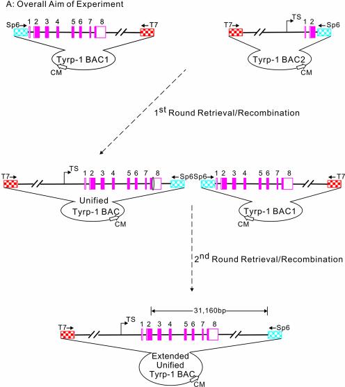
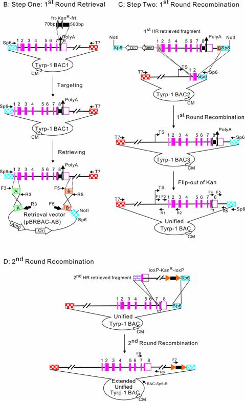
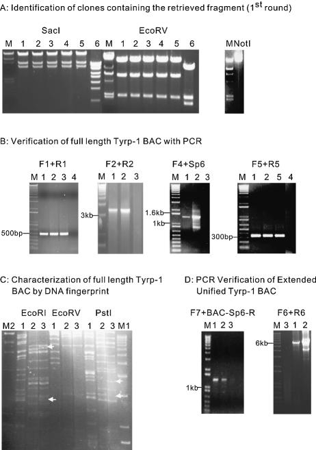
Recombinogenic engineering or recombineering is a powerful new method to engineer DNA without the need for restriction enzymes or ligases. We report here a general method for using recombineering to combine overlapping bacterial artificial chromosomes (BACs) to build larger, unified BACs. In order to test the feasibility of using recombineering to combine two large DNA fragments (>20 kb), we constructed a unified BAC containing the full-length tyrosinase-related protein-1 (Tyrp-1) gene from two library-derived BACs, one containing the 5' regulatory elements and the other containing the 3' coding exons. This was achieved using a two-step homologous recombination method enabled by the bacteriophage lambda Red proteins. In the first step, retrieval, a large DNA fragment (approximately 22 kb) was retrieved from one of the original BACs. In the second step, recombination, the retrieved DNA fragment was inserted into the second original BAC to form the unified BAC containing all the desired Tyrp-1 sequence. To further demonstrate the general applicability of our approach, an additional DNA fragment (approximately 20 kb) was inserted into the unified BAC downstream of the coding region. This method should prove very useful for enabling BAC manipulation in a variety of scenarios.


Figures

Figure 1
Figure 1
General experimental strategy for BAC engineering. The light blue diamond hatched pattern represents the Sp6 fragment of BAC. The red square checked pattern represents the T7 fragment of BAC. The green vertically striped pattern represents ArmA at the breakpoint of the Tyrp-1 gene of Tyrp-1 BAC1. The orange mosaic pattern represents ArmB at the end of the Tyrp-1 gene of BAC1. The pink solid rectangles represent exons of the Tyrp-1 gene. Double backslash lines represent disproportional genomic DNA. Amp is the ampicillin resistance gene. Ori refers to the origin of replication. The jointed arrow represents the transcriptional site; the vertical arrow represents the poly(A) site; the solid square with an oval on each side represents the 3749 bp frt–KanR–frt cassette (the oval represents the frt site); the solid rectangle with a triangle on each side represents 1896 bp of the loxP–KanR–loxP cassette. (A) Overall aim of the experiment. Top left: Tyrp-1 BAC1 containing Tyrp-1 exons 1–8 and the 3′ downstream region. Top right: Tyrp-1 BAC2 containing the Tyrp-1 5′ region and exons 1 and 2. Bottom: the final extended unified Tyrp-1 BAC containing the Tyrp-1 5′ region, all the exons and a 16.5 kb 3′ region. 31 160 bp is the distance between the 3′ breakpoint of Tyrp-1 BAC2 and the last base pair in the extended unified Tyrp-1 BAC. (B) Step one: first round retrieval (upper left). A kanamycin gene is first inserted into Tyrp-1 BAC1. The Tyrp-1 genomic DNA tagged by the kanamycin gene is then cloned into the retrieval vector (pBRBAC-AB). The positions of the primers which were used to amplify two homology arms are indicated by F3 and R3, and F5 and R5. In the retrieval vector, the ampicillin gene and origin of replication site are indicated by open arrows; the location of primers used to amplify the pBRBAC-AB retrieve vector are shown as thick closed arrows (R3 and F5). Homologous recombination events are denoted by crosses. NotI cleavage sites are also shown. (C) Step two: first round recombination (upper right). Linearized Tyrp-1 genomic fragment containing Tyrp-1 exons 1–8 and the 4 kb 3′ region was recombined with Tyrp-1 BAC2 through the homologous ArmA and Sp6 DNA regions. The resulting Tyrp-1 BAC3 and the unified Tyrp-1 BAC contained the 5′ regulatory region, all the Tyrp-1 exons and a 4 kb 3′ downstream region. The position and orientation of primers (F1, R1, F2, R2, F4, F5, Sp6 and R5) that were used to detect and sequence the BACs are shown. (D) Second round recombination (lower). A second round of retrieval–recombination was carried out with the unified Tyrp-1 BAC and a 19 891 bp fragment downstream of exon 8 to generate an extended unified Tyrp-1 BAC. The purple brick pattern represents a long homologous arm at Tyrp-1 intron 7 upstream of the frt site in the unified Tyrp-1 BAC. The positions and orientations of the primers (F6, F7, R6 and BAC-Sp6-R) that were used to detect the extended unified Tyrp-1 BAC are shown.
Figure 1
Figure 1
General experimental strategy for BAC engineering. The light blue diamond hatched pattern represents the Sp6 fragment of BAC. The red square checked pattern represents the T7 fragment of BAC. The green vertically striped pattern represents ArmA at the breakpoint of the Tyrp-1 gene of Tyrp-1 BAC1. The orange mosaic pattern represents ArmB at the end of the Tyrp-1 gene of BAC1. The pink solid rectangles represent exons of the Tyrp-1 gene. Double backslash lines represent disproportional genomic DNA. Amp is the ampicillin resistance gene. Ori refers to the origin of replication. The jointed arrow represents the transcriptional site; the vertical arrow represents the poly(A) site; the solid square with an oval on each side represents the 3749 bp frt–KanR–frt cassette (the oval represents the frt site); the solid rectangle with a triangle on each side represents 1896 bp of the loxP–KanR–loxP cassette. (A) Overall aim of the experiment. Top left: Tyrp-1 BAC1 containing Tyrp-1 exons 1–8 and the 3′ downstream region. Top right: Tyrp-1 BAC2 containing the Tyrp-1 5′ region and exons 1 and 2. Bottom: the final extended unified Tyrp-1 BAC containing the Tyrp-1 5′ region, all the exons and a 16.5 kb 3′ region. 31 160 bp is the distance between the 3′ breakpoint of Tyrp-1 BAC2 and the last base pair in the extended unified Tyrp-1 BAC. (B) Step one: first round retrieval (upper left). A kanamycin gene is first inserted into Tyrp-1 BAC1. The Tyrp-1 genomic DNA tagged by the kanamycin gene is then cloned into the retrieval vector (pBRBAC-AB). The positions of the primers which were used to amplify two homology arms are indicated by F3 and R3, and F5 and R5. In the retrieval vector, the ampicillin gene and origin of replication site are indicated by open arrows; the location of primers used to amplify the pBRBAC-AB retrieve vector are shown as thick closed arrows (R3 and F5). Homologous recombination events are denoted by crosses. NotI cleavage sites are also shown. (C) Step two: first round recombination (upper right). Linearized Tyrp-1 genomic fragment containing Tyrp-1 exons 1–8 and the 4 kb 3′ region was recombined with Tyrp-1 BAC2 through the homologous ArmA and Sp6 DNA regions. The resulting Tyrp-1 BAC3 and the unified Tyrp-1 BAC contained the 5′ regulatory region, all the Tyrp-1 exons and a 4 kb 3′ downstream region. The position and orientation of primers (F1, R1, F2, R2, F4, F5, Sp6 and R5) that were used to detect and sequence the BACs are shown. (D) Second round recombination (lower). A second round of retrieval–recombination was carried out with the unified Tyrp-1 BAC and a 19 891 bp fragment downstream of exon 8 to generate an extended unified Tyrp-1 BAC. The purple brick pattern represents a long homologous arm at Tyrp-1 intron 7 upstream of the frt site in the unified Tyrp-1 BAC. The positions and orientations of the primers (F6, F7, R6 and BAC-Sp6-R) that were used to detect the extended unified Tyrp-1 BAC are shown.
Figure 2
Figure 2
(A) Identification of clones containing the retrieved fragment (first round). The left panel shows the digestion pattern of the retrieved BAC fragment. M: λ/HindIII DNA. Different clones labeled 1–6 were digested by the restriction enzymes labeled above. Clones 1–5 show the expected digestion pattern. The right panel shows a 25.4 kb NotI-linearized pBRBAC-AB construct containing the 22.5 kb retrieved fragment. (B) Verification of Tyrp-1 BACs with PCR. A PCR product of 3.3 kb by primer pair (F2 + R2) could only be generated after homologous recombination occurred between the retrieved DNA and Tyrp-1 BAC2 to generate Tyrp-1 BAC3 and Tyrp-1 BAC. The four figures show the PCR verification of the full-length Tyrp-1 BAC3 and Tyrp-1 BAC with different primer pairs (F1 + R1, F2 + R2, F4 + Sp6 and F5 + R5). Each picture shows a band of the expected size. M: 1 kb plus Marker. Template DNAs used are indicated by numbers above each lane. (1) Tyrp-1 BAC3; (2) unified Tyrp-1 BAC; (3) Tyrp-1 BAC2; (4) PCR negative control (water); (5) Tyrp-1 BAC1. (C) Characterization of Tyrp-1 BACs by DNA fingerprint. The figure shows the DNA fingerprint results. The digestion pattern of unified Tyrp-1 BAC (labeled as 3) is very similar to that of the Tyrp-1 BAC2 (labeled as 2), but it also contains fragments unique to Tyrp-1 BAC1 (labeled as 1). The white arrows indicate DNA fragments present in unified Tyrp-1 BAC as well as in either Tyrp-1 BAC2 or Tyrp-1 BAC1, but not both. M1, λ/HindIII DNA; M2, 1 kb plus Marker. The restriction enzymes used are listed at the top of each panel. (D) PCR verification of second round recombination products. The two figures show the PCR verification of the second round recombination products. Two PCR products of 1.38 kb (left panel) and 6.2 kb (right panel) by primer pair (F7 + BAC-Sp6-R) and (F6 + R6), respectively, can only be generated after the second round recombination has occurred between the 19 891 bp retrieved DNA and Tyrp-1 BAC, creating the extended unified Tyrp-1 BAC. Each figure shows bands of the expected size. M: 1 kb plus Marker. Template DNAs used are indicated by numbers above each lane: lane 1, clone 1 of extended unified Tyrp-1 BAC; lane 2, clone 2 of extended unified Tyrp-1 BAC; lane 3, unified Tyrp-1 BAC.

Similar articles

Cited by

References

    1. Copeland N.G., Jenkins,N.A. and Court,D.L. (2001) Recombineering: a powerful new tool for mouse functional genomics. Nature Rev. Genet., 2, 769–779. - PubMed
    1. Muyrers J.P., Zhang,Y. and Stewart,A.F. (2001) Techniques: recombinogenic engineering—new options for cloning and manipulating DNA. Trends Biochem. Sci., 26, 325–331. - PubMed
    1. Cohen S.N., Chang,A.C., Boyer,H.W. and Helling,R.B. (1973) Construction of biologically functional bacterial plasmids in vitro. Proc. Natl Acad. Sci. USA, 70, 3240–3244. - PMC - PubMed
    1. Shizuya H., Birren,B., Kim,U.J., Mancino,V., Slepak,T., Tachiiri,Y. and Simon,M. (1992) Cloning and stable maintenance of 300-kilobase-pair fragments of human DNA in Escherichia coli using an F-factor-based vector. Proc. Natl Acad. Sci. USA, 89, 8794–8797. - PMC - PubMed
    1. Zhang Y., Buchholz,F., Muyrers,J.P.P. and Stewart,A.F. (1998) A new logic for DNA engineering using recombination in Escherichia coli. Nature Genet., 20, 123–128. - PubMed

Publication types