

Neural correlates of person recognition
- PMID: 12888543
- PMCID: PMC202315
- DOI: 10.1101/lm.57403
Neural correlates of person recognition
Abstract
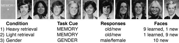
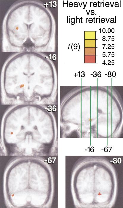
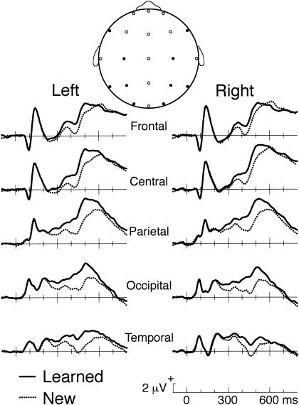
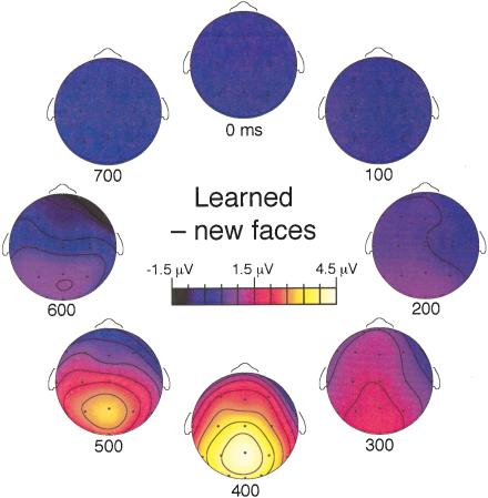
Rapidly identifying known individuals is an essential skill in human society. To elucidate the neural basis of this skill, we monitored brain activity while experimental participants demonstrated their ability to recognize people on the basis of viewing their faces. Each participant first memorized the faces of 20 individuals who were not known to the participants in advance. Each face was presented along with a voice simulating the individual speaking their name and a biographical fact. Following this learning procedure, the associated verbal information could be recalled accurately in response to each face. These learned faces were subsequently viewed together with new faces in a memory task. Subjects made a yes-no recognition decision in response to each face while also covertly retrieving the person-specific information associated with each learned face. Brain activity that accompanied this retrieval of person-specific information was contrasted to that when new faces were processed. Functional magnetic resonance imaging in 10 participants showed that several brain regions were activated during blocks of learned faces, including left hippocampus, left middle temporal gyrus, left insula, and bilateral cerebellum. Recordings of event-related brain potentials in 10 other participants tracked the time course of face processing and showed that learned faces engaged neural activity responsible for person recognition 300-600 msec after face onset. Collectively, these results suggest that the visual input of a recently learned face can rapidly trigger retrieval of associated person-specific information through reactivation of distributed cortical networks linked via hippocampal connections.
Figures
Comment in
-
Remembering people: neuroimaging takes on the real world.Learn Mem. 2003 Jul-Aug;10(4):240-1. doi: 10.1101/lm.65003. Learn Mem. 2003. PMID: 12888540 No abstract available.
References
-
- Aguirre, G.K., Singh, R., and D'Esposito, M. 1999. Stimulus inversion and the responses of face and object-sensitive cortical areas. Neuroreport 10: 189–194. - PubMed
-
- Allison, T., Ginter, H., McCarthy, G., Nobre, A.C., Puce, A., Luby, M., and Spencer, D.D. 1994. Face recognition in human extrastriate cortex. J. Neurophysiol. 71: 821–825. - PubMed
-
- Andreasen, N.C., O'Leary, D.S., Arndt, S., Cizadlo, T., Hurtig, R., Rezai, K., Watkins, G.L., Ponto, L.B., and Hichwa, R.D. 1996. Neural substrates of facial recognition. J. Neuropsychiat. Clin. Neurosci. 8: 139–146. - PubMed
-
- Beeman, M. 1998. Coarse semantic coding and discourse comprehension. In Right hemisphere language comprehension: Perspectives from cognitive neuroscience (eds. M. Beeman and C. Chiarello), pp. 255–284. Erlbaum, Mahwah, NJ.
Publication types
MeSH terms
Grants and funding
LinkOut - more resources
Full Text Sources
Medical