

Spared anterograde memory for shock-probe fear conditioning after inactivation of the amygdala
- PMID: 12888544
- PMCID: PMC202316
- DOI: 10.1101/lm.54103
Spared anterograde memory for shock-probe fear conditioning after inactivation of the amygdala
Abstract
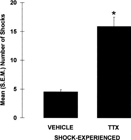
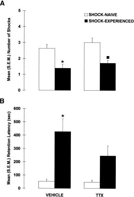
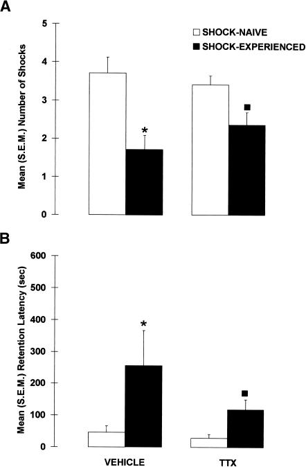
Previous studies have shown that amygdala lesions impair avoidance of an electrified probe. This finding has been interpreted as indicating that amygdala lesions reduce fear. It is unclear, however, whether amygdala-lesioned rats learn that the probe is associated with shock. If the lesions prevent the formation of this association, then pretraining reversible inactivation of the amygdala should impair both acquisition and retention performance. To test this hypothesis, the amygdala was inactivated (tetrodotoxin; TTX; 1 ng/side) before a shock-probe acquisition session, and retention was tested 4 d later. The data indicated that, compared with rats infused with vehicle, rats infused with TTX received more shocks during the acquisition session, but more importantly, were not impaired on the retention test. In Experiment 2, we assessed whether the spared memory on the retention test was caused by overtraining during acquisition. We used the same procedure as in Experiment 1, with the exception that the number of shocks the rats received during the acquisition session was limited to four. Again the data indicated that amygdala inactivation did not impair performance on the retention test. These results indicate that amygdala inactivation does not prevent the formation of an association between the shock and the probe and that shock-probe deficits during acquisition likely reflect the amygdala's involvement in other processes.
Figures
Similar articles
-
Amygdala lesions do not impair shock-probe avoidance retention performance.Behav Neurosci. 2000 Feb;114(1):107-16. doi: 10.1037//0735-7044.114.1.107. Behav Neurosci. 2000. PMID: 10718266
-
The lateral amygdala processes the value of conditioned and unconditioned aversive stimuli.Neuroscience. 2005;133(2):561-9. doi: 10.1016/j.neuroscience.2005.02.043. Neuroscience. 2005. PMID: 15878802
-
Context-dependent effects of hippocampal damage on memory in the shock-probe test.Hippocampus. 2005;15(1):18-25. doi: 10.1002/hipo.20024. Hippocampus. 2005. PMID: 15390168
-
Inactivation of the dorsal or ventral hippocampus with muscimol differentially affects fear and memory.Brain Res. 2010 Sep 24;1353:145-51. doi: 10.1016/j.brainres.2010.07.030. Epub 2010 Jul 18. Brain Res. 2010. PMID: 20647005
-
The amygdala, fear, and memory.Ann N Y Acad Sci. 2003 Apr;985:125-34. doi: 10.1111/j.1749-6632.2003.tb07077.x. Ann N Y Acad Sci. 2003. PMID: 12724154 Review.
Cited by
-
Effects of muscarinic receptor antagonism in the basolateral amygdala on two-way active avoidance.Exp Brain Res. 2011 Mar;209(3):455-64. doi: 10.1007/s00221-011-2576-4. Epub 2011 Feb 12. Exp Brain Res. 2011. PMID: 21318348
-
Posttraumatic stress disorder: a theoretical model of the hyperarousal subtype.Front Psychiatry. 2014 Apr 4;5:37. doi: 10.3389/fpsyt.2014.00037. eCollection 2014. Front Psychiatry. 2014. PMID: 24772094 Free PMC article. Review.
-
Basolateral amygdala lesions do not prevent memory of context-footshock training.Learn Mem. 2003 Nov-Dec;10(6):495-502. doi: 10.1101/lm.64003. Learn Mem. 2003. PMID: 14657260 Free PMC article.
References
-
- Ambrogi Lorenzini, C.G., Baldi, E., Bucherelli, C., Sacchetti, B., and Tassoni, G. 1997. Analysis of mnemonic processing by means of totally reversible neural inactivations. Brain Res. Brain Res. Protoc. 1: 391–398. - PubMed
-
- Antoniadis, E.A. and McDonald, R.J. 2001. Amygdala, hippocampus, and unconditioned fear. Exp. Brain Res. 138: 200–209. - PubMed
-
- Bast, T., Zhang, W.N., and Feldon, J. 2001. The ventral hippocampus and fear conditioning in rats. Different anterograde amnesias of fear after tetrodotoxin inactivation and infusion of the GABA(A) agonist muscimol. Exp. Brain Res. 139: 39–52. - PubMed
-
- Bermudez-Rattoni, F., Introini-Collison, I., Coleman-Mesches, K., and McGaugh, J.L. 1997. Insular cortex and amygdala lesions induced after aversive training impair retention: Effects of degree of training. Neurobiol. Learn. Mem. 67: 57–63. - PubMed
Publication types
MeSH terms
Substances
Grants and funding
LinkOut - more resources
Full Text Sources
Medical