Marginal biotin deficiency is teratogenic in ICR mice
- PMID: 12888630
- PMCID: PMC1450011
- DOI: 10.1093/jn/133.8.2519
Marginal biotin deficiency is teratogenic in ICR mice
Abstract
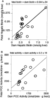
The incidence of marginal biotin deficiency in normal human gestation is approximately one in three. In ICR mice, maternal biotin deficiency results in cleft palate, micrognathia, microglossia and limb hypoplasia. However, the relationships among the severity of maternal biotin deficiency, fetal biotin status and malformations have not been reported. This study utilized validated indices of biotin status to investigate the relationships among maternal biotin status, fetal biotin status and the rate of fetal malformations in ICR mice. Biotin status was controlled by feeding diets with varying egg white concentration. In dams and fetuses, biotin status was assessed by hepatic biotin content and hepatic activity of the biotin-dependent enzyme propionyl-CoA carboxylase; in dams, status was also assessed by urinary excretion of biotin and 3-hydroxyisovaleric acid. Malformations were assessed morphologically. Biotin was measured by HPLC/avidin-binding assay. Propionyl-CoA carboxylase (PCC) activity was determined by H(14)CO(3) incorporation. 3-Hydroxyisovaleric acid concentration was determined by GC/MS. Although no overt signs of deficiency appeared, metabolic disturbances caused by biotin deficiency were detectable in dams and fetuses. These disturbances increased with increasing egg white. Fetal biotin status correlated significantly with maternal biotin status (fetal vs. dam hepatic biotin, r = 0.671; fetal vs. dam PCC activity, r = 0.70). The incidences of malformations were strikingly dependent on egg white concentration. We conclude that in ICR mice, marginal maternal biotin deficiency causes fetal biotin deficiency. We speculate that the fetal malformations are primarily the consequence of fetal biotin deficiency. Because murine malformations appeared at degrees of biotin deficiency that are similar to those in human gestation, we speculate that some human fetal malformations may be caused by biotin deficiency.
Figures


References
-
- Mock DM, Stadler DD. Conflicting indicators of biotin status from a cross-sectional study of normal pregnancy. J. Am. Coll. Nutr. 1997;16:252–257. - PubMed
-
- Mock DM, Stadler D, Stratton S, Mock NI. Biotin status assessed longitudinally in pregnant women. J. Nutr. 1997;127:710–716. - PubMed
-
- Zempleni J, Mock DM. Marginal biotin deficiency is teratogenic. Proc. Soc. Exp. Biol. Med. 2000;223:14–21. - PubMed
-
- Watanabe T, Endo A. Teratogenic effects of avidin-induced biotin deficiency in mice. Teratology. 1984;30:91–94. - PubMed
-
- Watanabe T. Teratogenic effects of biotin deficiency in mice. J. Nutr. 1983;113:574–581. - PubMed
Publication types
MeSH terms
Substances
Grants and funding
LinkOut - more resources
Full Text Sources
Miscellaneous
