. 2003 Aug;49(4):929-45.
doi: 10.1046/j.1365-2958.2003.03619.x.

Pathogenic Leptospira species express surface-exposed proteins belonging to the bacterial immunoglobulin superfamily

Affiliations

Pathogenic Leptospira species express surface-exposed proteins belonging to the bacterial immunoglobulin superfamily

James Matsunaga et al. Mol Microbiol. 2003 Aug.

Abstract

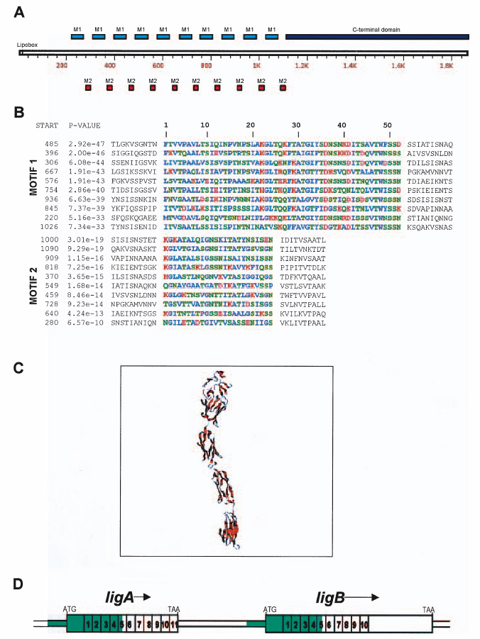
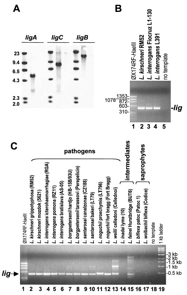
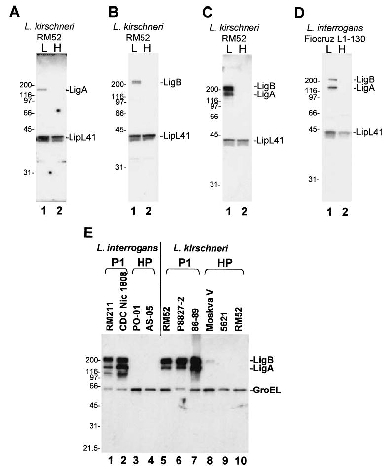
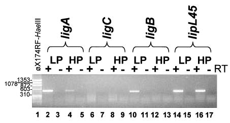
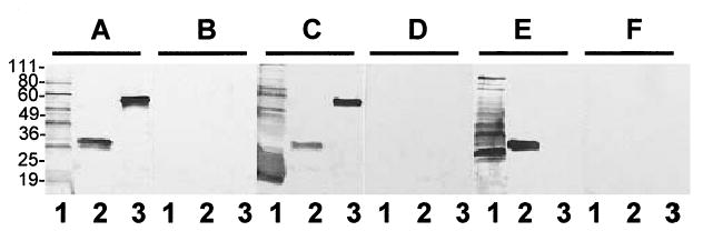
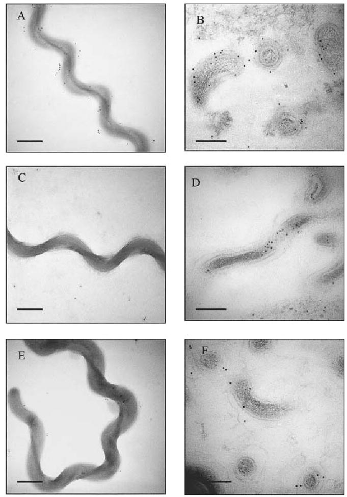
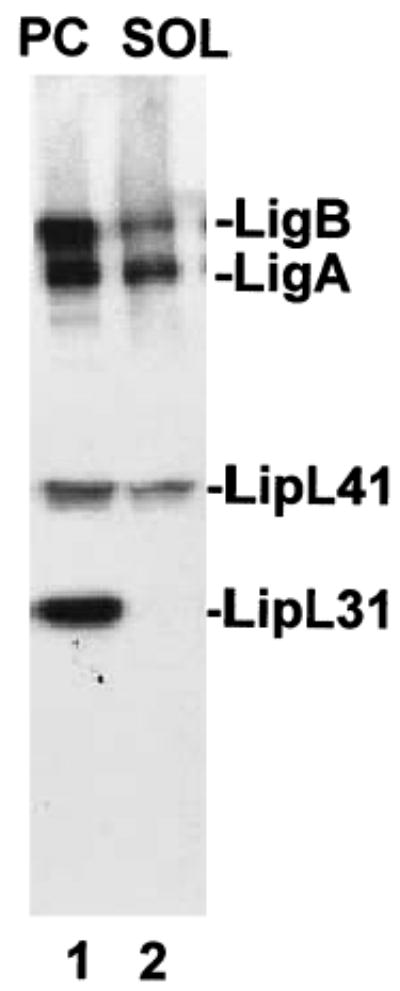
Proteins with bacterial immunoglobulin-like (Big) domains, such as the Yersinia pseudotuberculosis invasin and Escherichia coli intimin, are surface-expressed proteins that mediate host mammalian cell invasion or attachment. Here, we report the identification and characterization of a new family of Big domain proteins, referred to as Lig (leptospiral Ig-like) proteins, in pathogenic Leptospira. Screening of L. interrogans and L. kirschneri expression libraries with sera from leptospirosis patients identified 13 lambda phage clones that encode tandem repeats of the 90 amino acid Big domain. Two lig genes, designated ligA and ligB, and one pseudogene, ligC, were identified. The ligA and ligB genes encode amino-terminal lipoprotein signal peptides followed by 10 or 11 Big domain repeats and, in the case of ligB, a unique carboxy-terminal non-repeat domain. The organization of ligC is similar to that of ligB but contains mutations that disrupt the reading frame. The lig sequences are present in pathogenic but not saprophytic Leptospira species. LigA and LigB are expressed by a variety of virulent leptospiral strains. Loss of Lig protein and RNA transcript expression is correlated with the observed loss of virulence during culture attenuation of pathogenic strains. High-pressure freeze substitution followed by immunocytochemical electron microscopy confirmed that the Lig proteins were localized to the bacterial surface. Immunoblot studies with patient sera found that the Lig proteins are a major antigen recognized during the acute host infection. These observations demonstrate that the Lig proteins are a newly identified surface protein of pathogenic Leptospira, which by analogy to other bacterial immunoglobulin superfamily virulence factors, may play a role in host cell attachment and invasion during leptospiral pathogenesis.


Figures

Fig. 1
Fig. 1
A. Schematic representation of LigB. Motif 1 (blue box) and Motif 2 (red box) comprise the 10 Big2 repetitive domains from residue 220 through 1109, whereas the C-terminus (dark blue box) is located between 1111 and 1890. B. MEME Motif Discovery analysis of the Big2 repetitive domains of LigB. L. interrogans Lig proteins contain ~90mer repeats composed of two motifs of length 52 residues (M1) and 24 residues (M2), respectively. Residues are coloured based on their chemical properties: hydrophobic, basic, acidic and polar uncharged. The sites are listed in order of decreasing statistical significance (P-value). The P-values and start sites of the motifs are indicated. The P-value of a site is computed from the match score of the site with the position specific scoring matrix for the motif and represents the probability of a random string (generated from the background letter frequencies) having the same match score or higher. C. Comparative modelling of L. interrogans LigB repetitive domains (residues 493–917). D. Genetic structure of the ligAB locus. The 11 and 10 Big2 domains encoded by ligA and ligB, respectively, are numbered. Two additional N-terminal Big2 domains with poor E-values (>10−10) are not shown. The >2 kb segment of DNA sequence identity between the 5′ portions of ligA and ligB is indicated in green.
Fig. 2
Fig. 2
Detection of lig sequences in Leptospira species. A. Southern blot analysis. Genomic DNA from L. interrogans Fiocruz L1-130 (middle lane) and L. biflexa Patoc 1 (right lane) were digested with NsiI and PacI and probed in Southern blots with ligA, ligB and ligC specific probes. B and C. PCR analysis. Leptospiral DNA was subjected to PCR analysis by degenerate primers designed to anneal to all lig genes in L. kirschneri RM52 and L. interrogans Fiocruz L1-130. The expected size of the PCR product is −0.5 kb.
Fig. 3
Fig. 3
Western blot analysis of LigA and LigB expression in low- and high-passage Leptospira. A and B. Immunoblots of low (9 passages) and high-passage (>200 passages) isolates of L. kirschneri RM52 were probed with LipL41 antiserum (1:10 000), and either LigA antiserum (A, 1:2000) or LigB antiserum (B, 1:2000). L, low passage; H, high passage. C and D. Immunoblots of L. kirschneri RM52 or L. interrogans Fiocruz L1-130 (passage 25 and passage 69) were probed with LipL41 (1:10 000) and LigA/B repeat antisera (1:5000). E. An immunoblot of L. interrogans and L. kirschneri isolates was probed with LigA/B repeat antisera (1:5000) and GroEL antisera (1:30 000). Strain names are shown above each lane. P1, passage 1; HP, high passage. The relative mobility (kDa) of molecular mass standards (Bio-Rad) is shown on the left of each panel.
Fig. 4
Fig. 4
RT-PCR analysis of lig transcripts in low- and high-passage L. kirschneri RM52. Total RNA from low- (passage 4) and high-passage L. kirschneri RM52 was analysed by RT-PCR with primers specific for each lig gene. +, reverse transcriptase present in the reaction; −, reverse transcriptase omitted.
Fig. 5
Fig. 5
Reactivity of sera from leptospirosis patients and Rattus norvegicus reservoirs with recombinant LigB. Membranes were prepared from SDS-PAGE of whole L. interrogans extract (108 organisms per lane, lanes 1); recombinant L. interrogans lipoprotein, LipL32 (1 μg per lane, lanes 2) and recombinant protein containing repeat domains 1–5 of L. interrogans LigB (1 μg per lane, lanes 3). Membranes were probed with sera from leptospirosis patients (A); healthy individuals (B); captured R. norvegicus from which L. interrogans was isolated (C); captured culture-negative R. norvegicus (D); laboratory rats immunized with whole-cell preparations of in vitro cultured low-passage L. interrogans (E); and laboratory rats obtained prior to immunization (F). The relative mobility (kDa) of molecular mass standards (Invitrogen) is shown on the left.
Fig. 6
Fig. 6
Immunoelectron microscopy of whole-cell (A, C and E) and thin-section (B, D, F) preparations of L. kirschneri strain RM52. Preparations were incubated with anti-LPS (A, B), anti-GroEL (C, D), and anti-LigB antibody (E, F), followed by anti-rabbit secondary antibody conjugated to 10 nm gold particles. Bars represent 100 nm.
Fig. 7
Fig. 7
Triton X-100 extraction of Lig proteins from L. kirschneri RM52. Leptospires were incubated for 2 h in MEM (ATCC) at 37°C. Bacteria were then washed once in 1× PBS with 5 mM MgCl2 and extracted with 1% Triton X-100. The insoluble protoplasmic cylinder (PC) was pelleted by centrifugation. The protoplasmic cylinder and the Triton-soluble (SOL) fractions were probed in a Western blot using LigA/B repeat (1:5000), LipL41 (1:10 000), and LipL31 (1:8000) antisera. LipL31 is a marker for the leptospiral inner membrane (Haake and Matsunaga, 2002).

References

    1. Arean VM. The pathologic anatomy and pathogenesis of fatal human leptospirosis (Weil’s disease) Am J Pathol. 1962;40:393–423. - PMC - PubMed
    1. Bailey TL, Elkan C. Fitting a mixture model by expectation maximization to discover motifs in biopolymers. Proc Int Conf Intell Syst Mol Biol. 1994;2:28–36. - PubMed
    1. Ballard SA, Williamson M, Adler B, Vinh T, Faine S. Interactions of virulent and avirulent leptospires with primary cultures of renal epithelial cells. J Med Microbiol. 1986;21:59–67. - PubMed
    1. Ballard SA, Segers RPAM, Bleumink-Pluym N, Fyfe J, Faine S, Adler B. Molecular analysis of the hsp (groE) operon of Leptospira interrogans serovar copenhageni. Mol Microbiol. 1993;8:739–751. - PubMed
    1. Ballard SA, Go M, Segers RP, Adler B. Molecular analysis of the dnaK locus of Leptospira interrogans serovar copenhageni. Gene. 1998;216:21–29. - PubMed

Publication types

MeSH terms

Associated data