

Protective effects of curcumin against amiodarone-induced pulmonary fibrosis in rats
- PMID: 12890714
- PMCID: PMC1573957
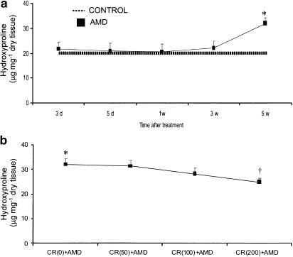
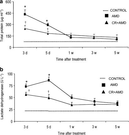
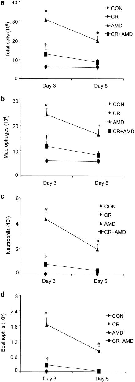
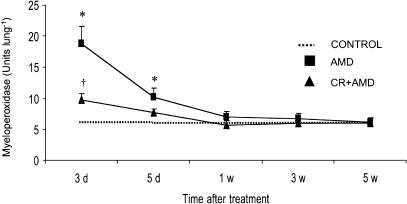
- DOI: 10.1038/sj.bjp.0705362
Protective effects of curcumin against amiodarone-induced pulmonary fibrosis in rats
Abstract
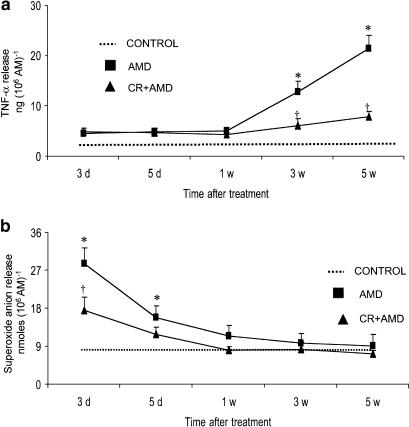
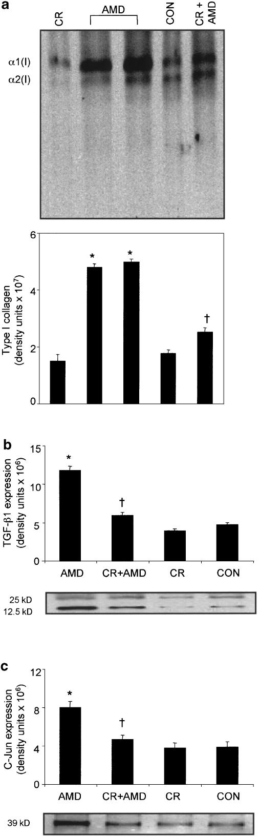
(1) We have studied whether curcumin prevents amiodarone-induced lung fibrosis in rats. Intratracheal instillation of amiodarone (6.25 mg kg(-1) on days 0 and 2, and then killed on day 3, day 5, week 1, week 3 and week 5 after amiodarone administration) induced increases in total protein and lactate dehydrogenase (LDH) activity on days 3 and 5 in bronchoalveolar lavage fluid (BALF). Total cell counts, alveolar macrophages, neutrophils and eosinophils recovered by BAL, and lung myeloperoxidase (MPO) activity were significantly higher in amiodarone rats. (2) Tumor necrosis factor-alpha (TNF-alpha) release after lipopolysaccharide (LPS) stimulation and superoxide anion generation after phorbol myristate acetate (PMA) stimulation were higher in the alveolar macrophages of amiodarone rats at 3 and 5 weeks postamiodarone instillation than in controls. Amiodarone also induced increases in transforming growth factor-beta1 (TGF-beta1) expression, collagen deposition, type I collagen expression and c-Jun protein in lungs. (3) Curcumin (200 mg kg(-1) body weight after first amiodarone instillation and daily thereafter for 5 weeks)-treated amiodarone rats had reduced levels of protein, LDH activity, total cell numbers and differential cell counts in BALF. LPS-stimulated TNF-alpha release and PMA-stimulated superoxide generation were significantly suppressed by curcumin. Furthermore, curcumin inhibited the increases in lung MPO activity, TGF-beta1 expression, lung hydroxyproline content, expression of type I collagen and c-Jun protein in amiodarone rats. Our results have important implications for the treatment of amiodarone-induced lung fibrosis.
Figures
Similar articles
-
[Effects of andrographolide on the concentration of cytokines in BALF and the expressions of type I and III collagen mRNA in lung tissue in bleomycin-induced rat pulmonary fibrosis].Xi Bao Yu Fen Zi Mian Yi Xue Za Zhi. 2011 Jul;27(7):725-9. Xi Bao Yu Fen Zi Mian Yi Xue Za Zhi. 2011. PMID: 21722520 Chinese.
-
Alveolar macrophage cytokine and growth factor production in a rat model of crocidolite-induced pulmonary inflammation and fibrosis.J Toxicol Environ Health. 1995 Oct;46(2):155-69. doi: 10.1080/15287399509532026. J Toxicol Environ Health. 1995. PMID: 7563215
-
Curcumin inhibition of bleomycin-induced pulmonary fibrosis in rats.Br J Pharmacol. 2000 Sep;131(2):169-72. doi: 10.1038/sj.bjp.0703578. Br J Pharmacol. 2000. PMID: 10991907 Free PMC article.
-
Amiodarone pulmonary toxicity: morphologic and biochemical features.Proc Soc Exp Biol Med. 1991 Jan;196(1):1-7. doi: 10.3181/00379727-196-43158a. Proc Soc Exp Biol Med. 1991. PMID: 1984236 Review. No abstract available.
-
Cytotoxicity and cytoprotective activities of natural compounds. The case of curcumin.Xenobiotica. 1996 Jul;26(7):667-80. doi: 10.3109/00498259609046741. Xenobiotica. 1996. PMID: 8819298 Review. No abstract available.
Cited by
-
Hydromorphone Protects against CO2 Pneumoperitoneum-Induced Lung Injury via Heme Oxygenase-1-Regulated Mitochondrial Dynamics.Oxid Med Cell Longev. 2021 Apr 9;2021:9034376. doi: 10.1155/2021/9034376. eCollection 2021. Oxid Med Cell Longev. 2021. PMID: 33927798 Free PMC article.
-
Impacts of Curcumin Treatment on Experimental Sepsis: A Systematic Review.Oxid Med Cell Longev. 2023 Jan 30;2023:2252213. doi: 10.1155/2023/2252213. eCollection 2023. Oxid Med Cell Longev. 2023. PMID: 36756300 Free PMC article.
-
Curcumin modulates the inflammatory response and inhibits subsequent fibrosis in a mouse model of viral-induced acute respiratory distress syndrome.PLoS One. 2013;8(2):e57285. doi: 10.1371/journal.pone.0057285. Epub 2013 Feb 20. PLoS One. 2013. PMID: 23437361 Free PMC article.
-
Non-pharmacological treatment of Helicobacter pylori.World J Gastrointest Pharmacol Ther. 2016 May 6;7(2):171-8. doi: 10.4292/wjgpt.v7.i2.171. World J Gastrointest Pharmacol Ther. 2016. PMID: 27158532 Free PMC article. Review.
-
Targeting Oxidative Stress as a Therapeutic Approach for Idiopathic Pulmonary Fibrosis.Front Pharmacol. 2022 Jan 21;12:794997. doi: 10.3389/fphar.2021.794997. eCollection 2021. Front Pharmacol. 2022. PMID: 35126133 Free PMC article. Review.
References
-
- ABE Y., HASHIMOTO S., HORIE T. Curcumin inhibition of inflammatory cytokine production by human peripheral blood monocytes and alveolar macrophages. Pharmacol. Res. 1999;39:41–47. - PubMed
-
- AHSAN H., PARVEEN N., KHAN N.U., HADI S.M. Pro-oxidant, anti-oxidant and cleavage activities on DNA of curcumin and its derivatives demethoxycurcumin and bisdemethoxycurcumin. Chem. Biol. Interact. 1999;121:161–175. - PubMed
-
- AMMON H.P.T., WAHL M.A. Pharmacology of Curcuma longa. Planta. Med. 1991;57:1–7. - PubMed
-
- ANGEL P., KARIN M. The role of Jun, Fos and the AP-1 complex in cell-proliferation and transformation. Biochim. Biophys. Acta. 1991;1072:129–157. - PubMed
-
- ARAUJO C.A.C., LEON L.L. Biological activities of Curcuma longa L. Mem. Inst. Oswaldo Cruz. 2001;96:723–728. - PubMed
Publication types
MeSH terms
Substances
LinkOut - more resources
Full Text Sources
Medical
Research Materials
Miscellaneous
