

Analysis of the tumour suppressor genes, FHIT and WT-1, and the tumour rejection genes, BAGE, GAGE-1/2, HAGE, MAGE-1, and MAGE-3, in benign and malignant neoplasms of the salivary glands
- PMID: 12890744
- PMCID: PMC1187325
- DOI: 10.1136/mp.56.4.226
Analysis of the tumour suppressor genes, FHIT and WT-1, and the tumour rejection genes, BAGE, GAGE-1/2, HAGE, MAGE-1, and MAGE-3, in benign and malignant neoplasms of the salivary glands
Abstract
Aims: Molecular genetic changes involved in tumorigenesis and malignant transformation of human tumours are novel targets of cancer diagnosis and treatment. This study aimed to analyse the expression of putative tumour suppressor genes, FHIT and WT-1, and tumour rejection genes, BAGE, GAGE-1/2, MAGE-1, MAGE-3, and HAGE (which are reported to be important in human cancers), in salivary gland neoplasms.
Methods: Gene expression was analysed by reverse transcription polymerase chain reaction (RT-PCR) in normal salivary gland tissue and 44 benign and malignant salivary gland tumours.
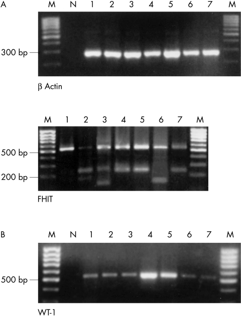
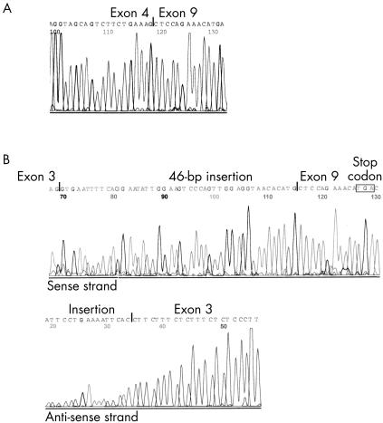
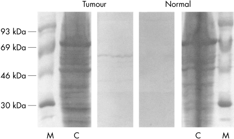
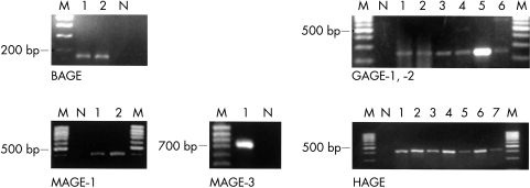
Results: Aberrant FHIT transcripts were found in one of 38 normal salivary glands, three of 28 adenomas, and two of 16 carcinomas. WT-1 mRNA was detectable in two adenomas and five carcinomas. Immunoblotting showed that WT-1 mRNA expression was associated with raised WT-1 protein concentrations. RT-PCR for detection of BAGE, GAGE, and MAGE gene expression was positive in two adenomas and nine carcinomas, but negative in normal salivary gland tissue. HAGE mRNA was found in two normal salivary glands, 11 benign, and eight malignant tumours.
Conclusions: FHIT mRNA splicing does not appear to be involved in the genesis of salivary gland neoplasms. The upregulation of WT-1 mRNA in tumours of epithelial/myoepithelial phenotype may imply a potential role of WT-1 in the genesis and/or cellular differentiation of these salivary gland tumours. The tumour rejection genes were more frequently, but not exclusively, expressed in malignant salivary gland tumours than in benign neoplasms, although none was suitable as a diagnostic marker of malignancy in salivary gland neoplasms.
Figures
References
-
- Ohta M, Inoue H, Cotticelli MG, et al. The FHIT gene, spanning the chromosome 3p14.2 fragile site and renal carcinoma-associated t(3;8) breakpoint, is abnormal in digestive tract cancers. Cell 1996;84:587–97. - PubMed
-
- Biéche I, Latil A, Becette V, et al. Study of FHIT transcripts in normal and malignant breast tissue. Genes Chromosomes Cancer 1998;23:292–9. - PubMed
-
- Bullerdiek J, Takla G, Barnitzke S, et al. Relationship of cytogenetic subtypes of salivary gland pleomorphic adenomas with patient age and histologic type. Cancer 1989;64:876–80. - PubMed
-
- Geurts JMW, Schoenmakers EFPM, Röijer E, et al. Expression of reciprocal hybrid transcripts of HMGIC and FHIT in a pleomorphic adenoma of the parotid gland. Cancer Res 1997;57:13–17. - PubMed
MeSH terms
Substances
LinkOut - more resources
Full Text Sources