. 2003 Jul 30;23(17):6778-87.
doi: 10.1523/JNEUROSCI.23-17-06778.2003.

The relay of high-frequency sensory signals in the Whisker-to-barreloid pathway

Affiliations

The relay of high-frequency sensory signals in the Whisker-to-barreloid pathway

Martin Deschênes et al. J Neurosci. .

Abstract

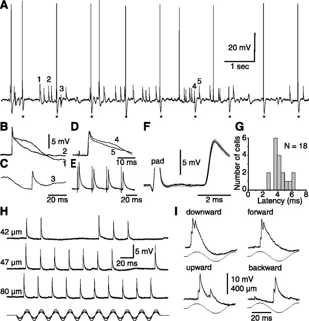
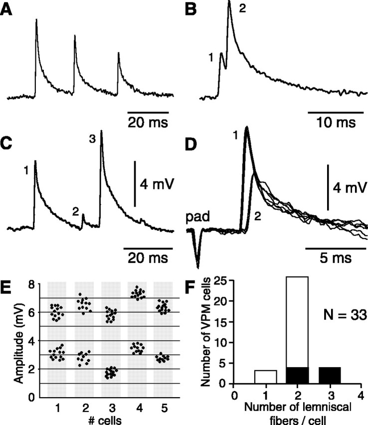
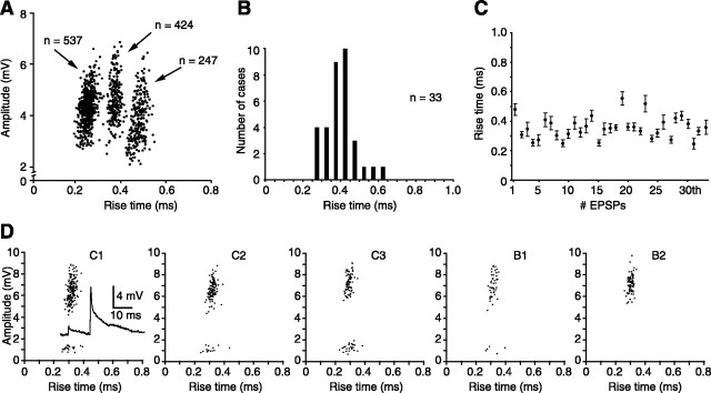
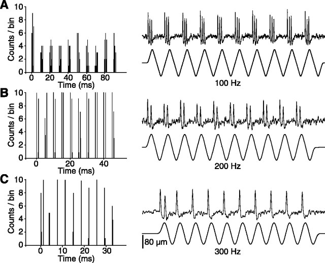
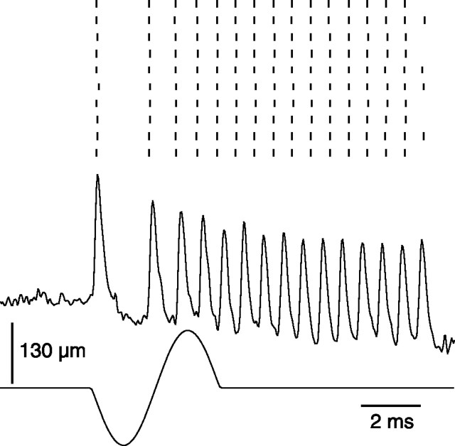
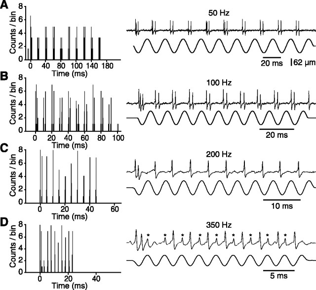
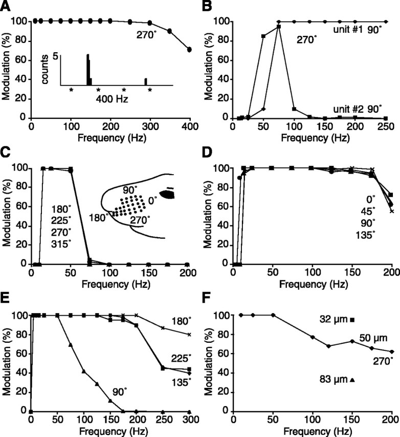
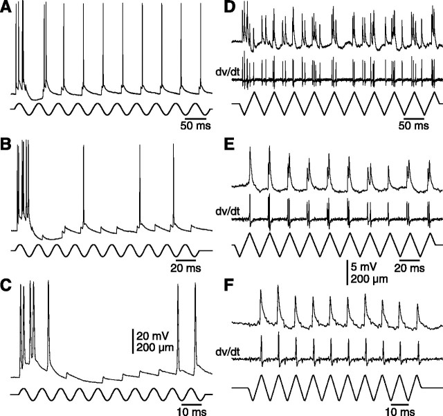
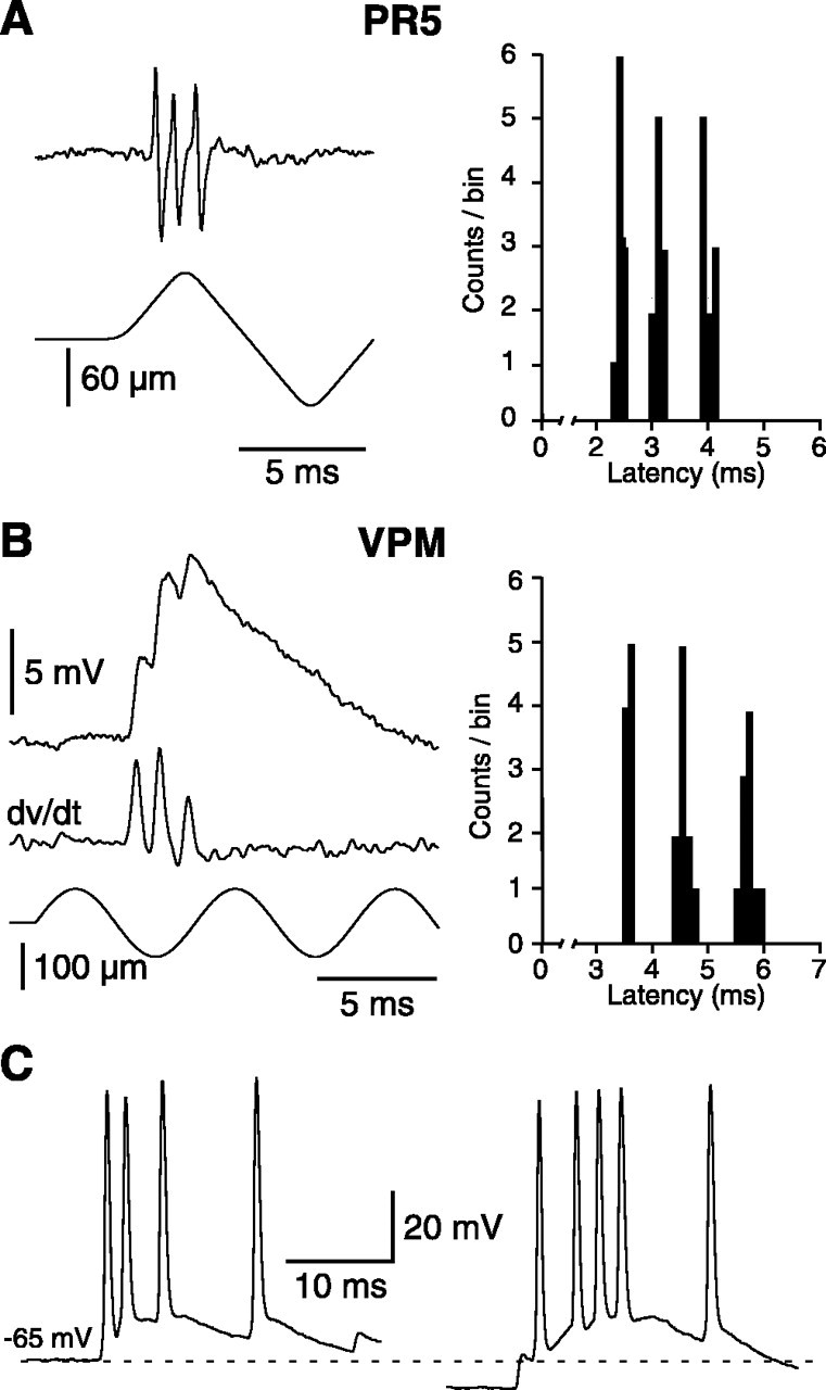
The present study investigated the operational features of whisker-evoked EPSPs in barreloid cells and the ability of the whisker-to-barreloid pathway to relay high rates of whisker deflection in lightly anesthetized rats. Results show that lemniscal EPSPs are single-fiber events with fast rise times (<500 microsec) that strongly depress at short inter-EPSP intervals. They occur at short latencies (3.84 +/- 0.96 msec) with little jitters (<300 microsec) after electrical stimulation of the whisker follicle. Waveform analysis indicates that one to three lemniscal axons converge on individual barreloid cells to produce EPSPs of similar rise times but different amplitudes. When challenged by high rates of whisker deflection, cells in the whisker-to-barreloid pathway demonstrate a remarkable frequency-following ability. Primary vibrissa afferents could follow in a phase-locked manner trains of sinusoidal deflections at up to 1 kHz. Although trigeminothalamic cells could still faithfully follow deflection rates of 200-300 Hz, the actual frequency-following ability of individual cells depends on the amplitude, velocity, and direction of displacements. The discharges of trigeminothalamic cells induce corresponding phase-locked EPSPs in barreloid cells, which trigger burst discharges at stimulus onset. During the following cycles of the stimulus train, few action potentials ensue because of the strong synaptic depression at lemniscal synapses. It is concluded that the whisker-to-barreloid pathway can relay vibratory inputs with a high degree of temporal precision, but that the relay of this information to the cerebral cortex requires the action of modulators, and possibly phase-locked discharges among an ensemble of relay cells.


Figures

Figure 1.
Figure 1.
Mechanical properties of the piezoelectric stimulator used for high-frequency whisker deflection. A, The probe consists of a straw attached to the bimorph bender. The cut tip of the vibrissa was inserted in the cone-shaped glass bead glued in the free end of the straw. B, The frequency-following property of the probe was monitored with a photodiode (pd) (deflection amplitude, 100 μm; stimulus frequency, 175 Hz). Note the notch (C, arrow) at stimulus onset and the ringing at stimulus offset (D) that reflect the resonance frequency of the probe (∼1 kHz).
Figure 2.
Figure 2.
Unitary character of whisker-evoked EPSPs in barreloid cells. A, Trace shows a short episode of spontaneously occurring and electrically evoked EPSPs (asterisks, follicular stimulation) in a D2-responsive relay cell. Events labeled 1-4 in A are magnified in B-D. Superimposed traces (B, D) show that EPSPs have similar rise times but variable falling phases. Note that the late component is much reduced when EPSPs are superimposed onto an IPSP (C). Time scale in C also applies to B. The thick trace in D is the stimulus-evoked response. E, Trace shows the depression of lemniscal EPSPs evoked in a C3-responsive cell by repetitive electrical stimulation of the whisker follicle. F, Superimposed traces (n = 10) of EPSPs evoked in a C2-responsive cell by suprathreshold electrical stimulation of the follicle (pad). Note the very small jitters of the bisynaptic responses. G, Shown are the shortest latencies of EPSPs evoked in 18 VPM cells by electrical stimulation of the whisker follicles. H, All-or-none occurrence of EPSPs evoked in a C2-responsive cell by straddling threshold (42 and 47 μm) and suprathreshold deflections (80 μm). I, Bursts of unitary EPSPs evoked at stimulus onset by deflecting whisker B4 in different directions. All of the cells but the one in I were classified as single-whisker neurons.
Figure 4.
Figure 4.
Criteria used to estimate the number of lemniscal fibers that contact single barreloid cells. A, Trace shows the synaptic depression observed when whisker-evoked EPSPs occurred at short intervals. In this case, it was inferred that the EPSP sequence resulted from the firing of a single presynaptic fiber. B, C, Traces show cases that break the rule of synaptic depression. To generate the sequences shown in B and C, two and three presynaptic fibers are required, respectively. D, Additional evidence for the convergence of lemniscal fibers on the same barreloid cells was obtained after electrical stimulation of the follicle (pad) or of the medial lemniscus. Superimposed traces (n = 6) show single-fiber EPSPs of different amplitudes evoked at slightly different stimulus intensities. E, Scatter plots summarize the amplitude distribution of evoked EPSPs in five additional cells (each plot contains 20-30 measures). F, On the basis of these criteria, the histogram shows the number of lemniscal fibers that contact each of the 33 barreloid cells analyzed. Filled bars represent cells that were strongly driven by multiple whiskers, and open bars represent single-whisker units.
Figure 3.
Figure 3.
Amplitude and rise time characteristics of whisker-evoked EPSPs in barreloid cells. A, Scatter plots show the relationship between the amplitudes and rise times (10-90% of peak amplitude) of EPSPs evoked by principal whisker deflection in three barreloid cells. The number of EPSPs in each cluster is indicated by arrows. B, The distribution of rise times across a population of 33 cells is shown. C, Graph shows the small dispersion of rise time values (error bars, SD). D, Plots show the relationship between the amplitudes and rise times of EPSPs evoked in a multiwhisker cell by deflecting separately each whisker composing its receptive field (whiskers are identified above the plots). Note the presence of at least two EPSPs of different amplitude (inset in plot C1), and their similar rise time values.
Figure 5.
Figure 5.
Frequency-following property of primary vibrissa afferents. The left-hand panels show PSTHs of cell responses to the stimulus sequences displayed in the right-hand panels. Each PSTH compiles 10 sequences.
Figure 6.
Figure 6.
Encoding of high-frequency whisker deflections by primary afferent axons. This B2-responsive unit fired in a one-to-one manner (middle trace) in phase with the resonance frequency of the stimulator (e.g., ∼1 kHz) (Fig. 1). Note the remarkable time locking of the discharges in the raster display.
Figure 7.
Figure 7.
Frequency-following property of PR5 cells to repetitive whisker deflections. The left-hand panels show PSTHs of cell discharges to the stimulus sequences displayed in the right-hand panels. Each PSTH compiles 10 sequences. Asterisks indicate induction transients picked up by the recording probe.
Figure 8.
Figure 8.
Representative examples of the ability of PR5 cells to follow high-frequency whisker stimulation. Cells were driven by bursts of 10 cycle sinusoidal displacements in different directions (0°, caudalward; 90°, upward, etc.) as shown in the inset in C. All of the units responded with at least one spike to the first cycle of each stimulus sequence. The percentage of modulation was computed by averaging the number of times the unit responded with at least one spike to each of the subsequent 9 cycles across a series of 10 stimulus sequences. The inserted PSTH in A shows responses evoked by electrical stimulation of the follicle (asterisks, 4 shocks at 400 Hz). See Results for full description.
Figure 9.
Figure 9.
Patterns of lemniscal EPSPs evoked in barreloid cells by repetitive whisker deflections. A-C, Traces show responses of a C2-responsive barreloid cell to bursts of sinusoidal deflections at 20, 50, and 100 Hz, respectively. D-F, Traces show EPSPs and their time derivatives recorded in another cell driven by bursts of triangular deflections. The cell was kept hyperpolarized to prevent spike discharges. Note that, at 20 Hz (D), EPSPs were evoked during both phases of the stimulus sequence (here, downward and upward deflections of vibrissa C4), but that, at higher rates of stimulation (50 and 100 Hz in E and F, respectively), EPSPs only followed upward deflections.
Figure 10.
Figure 10.
High firing rates induced in PR5 and barreloid cells by whisker deflections. A, Most PR5 units could generate stereotyped bursts of action potentials (intraburst frequencies, >1 kHz) in response to high-velocity whisker displacements. B, In barreloid cells, PR5 bursts induced compound EPSPs with similar intraburst frequencies. A, B, PSTHs show spikes (A) and EPSP counts (B) (discriminated by the time derivative of EPSPs) evoked by the first deflections of 10 stimulus sequences. C, The left-hand trace shows a burst driven by lemniscal EPSPs in response to the first cycle of a 100 Hz stimulus sequence. The right-hand trace shows the response to the same stimulus during membrane hyperpolarization. The dotted line indicates the resting potential (-65 mV).

Similar articles

Cited by

References

    1. Ahissar E, Sosnik R, Haidarliu S ( 2000) Transformation from temporal to rate coding in a somatosensory thalamocortical pathway. Nature 406: 302-306. - PubMed
    1. Alonso JM, Usrey WM, Reid RC ( 1996) Precisely correlated firing in cells of the lateral geniculate nucleus. Nature 383: 815-819. - PubMed
    1. Bae YC, Ihn HJ, Park MJ, Otterson OP, Moritani M, Yoshida A, Yoshio S ( 2000) Identification of signal substances in synapses made between primary afferents and their associated axon terminals in the rat trigeminal sensory nuclei. J Comp Neurol 418: 299-309. - PubMed
    1. Bloomfield SA, Sherman MS ( 1988) Postsynaptic potentials recorded in neurons of the cat's lateral geniculate nucleus following electrical stimulation of the optic chiasm. J Neurophysiol 60: 1924-1945. - PubMed
    1. Brecht M, Sakmann B ( 2002) Whisker maps of neuronal subclasses of the rat ventral posterior medial thalamus, identified by whole-cell voltage recording and morphological reconstruction. J Physiol (Lond) 538: 495-515. - PMC - PubMed

Publication types

MeSH terms

LinkOut - more resources