. 2003 Jul 30;23(17):6826-36.
doi: 10.1523/JNEUROSCI.23-17-06826.2003.

Enhanced expression of a specific hyperpolarization-activated cyclic nucleotide-gated cation channel (HCN) in surviving dentate gyrus granule cells of human and experimental epileptic hippocampus

Affiliations

Enhanced expression of a specific hyperpolarization-activated cyclic nucleotide-gated cation channel (HCN) in surviving dentate gyrus granule cells of human and experimental epileptic hippocampus

Roland A Bender et al. J Neurosci. .

Abstract

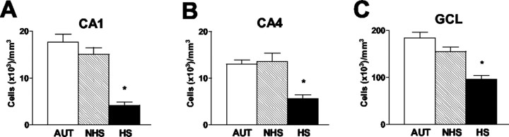
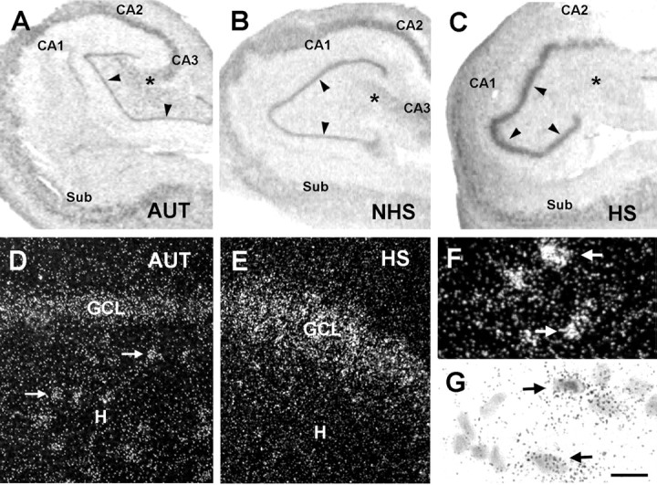
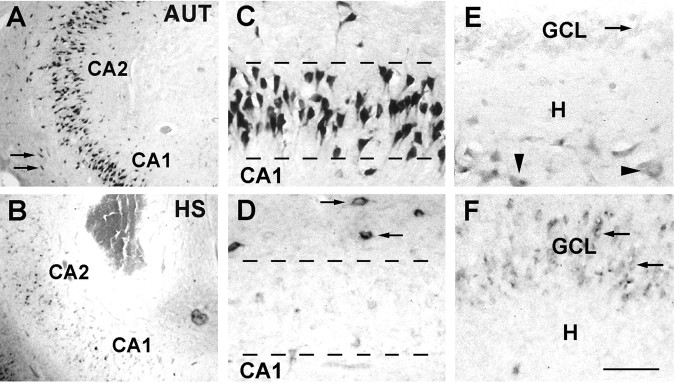
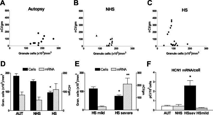
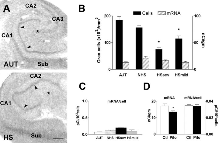
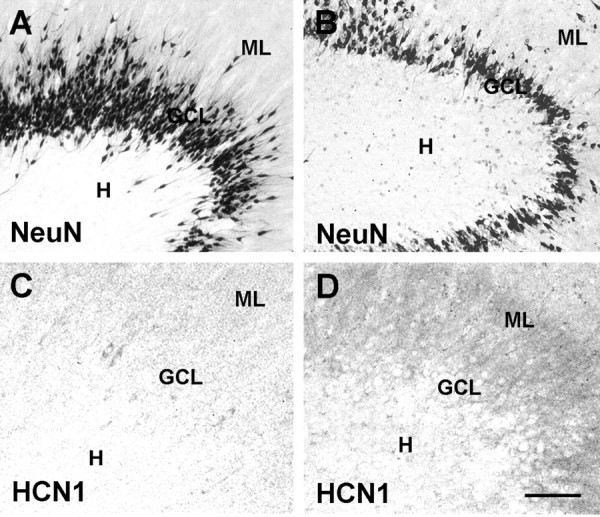
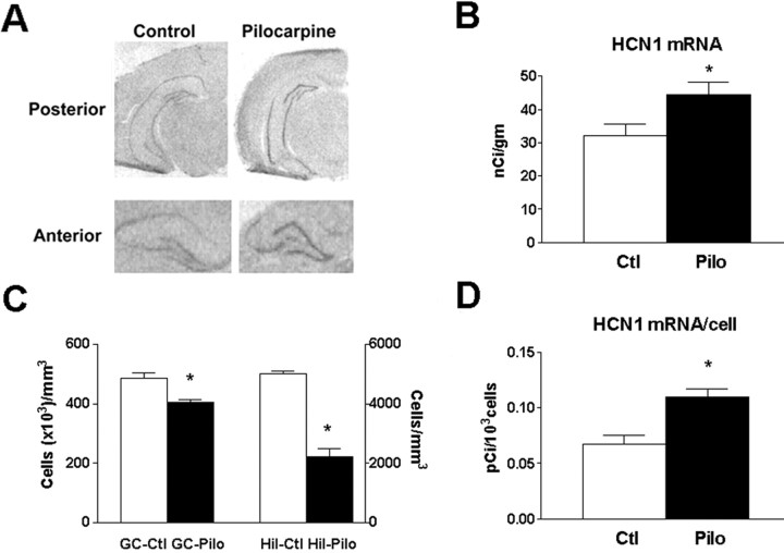
Changes in the expression of ion channels, contributing to altered neuronal excitability, are emerging as possible mechanisms in the development of certain human epilepsies. In previous immature rodent studies of experimental prolonged febrile seizures, isoform-specific changes in the expression of hyperpolarization-activated cyclic nucleotide-gated cation channels (HCNs) correlated with long-lasting hippocampal hyperexcitability and enhanced seizure susceptibility. Prolonged early-life seizures commonly precede human temporal lobe epilepsy (TLE), suggesting that transcriptional dysregulation of HCNs might contribute to the epileptogenic process. Therefore, we determined whether HCN isoform expression was modified in hippocampi of individuals with TLE. HCN1 and HCN2 expression were measured using in situ hybridization and immunocytochemistry in hippocampi from three groups: TLE with hippocampal sclerosis (HS; n = 17), epileptic hippocampi without HS, or non-HS (NHS; n = 10), and autopsy material (n = 10). The results obtained in chronic human epilepsy were validated by examining hippocampi from the pilocarpine model of chronic TLE. In autopsy and most NHS hippocampi, HCN1 mRNA expression was substantial in pyramidal cell layers and lower in dentate gyrus granule cells (GCs). In contrast, HCN1 mRNA expression over the GC layer and in individual GCs from epileptic hippocampus was markedly increased once GC neuronal density was reduced by >50%. HCN1 mRNA changes were accompanied by enhanced immunoreactivity in the GC dendritic fields and more modest changes in HCN2 mRNA expression. Furthermore, similar robust and isoform-selective augmentation of HCN1 mRNA expression was evident also in the pilocarpine animal model of TLE. These findings indicate that the expression of HCN isoforms is dynamically regulated in human as well as in experimental hippocampal epilepsy. After experimental febrile seizures (i.e., early in the epileptogenic process), the preserved and augmented inhibition onto principal cells may lead to reduced HCN1 expression. In contrast, in chronic epileptic HS hippocampus studied here, the profound loss of interneuronal and principal cell populations and consequent reduced inhibition, coupled with increased dendritic excitation of surviving GCs, might provoke a "compensatory" enhancement of HCN1 mRNA and protein expression.


Figures

Figure 1.
Figure 1.
Neuronal densities in hippocampal fields involved in the neuropathology of hippocampal sclerosis. A, CA1. B, CA4 (corresponding to CA3c in the rodent). C, GCL of the dentate gyrus. Neuronal counts and analyses were performed as described in Materials and Methods. As evident from the figure, whereas specimens from epileptic individuals without hippocampal sclerosis (NHS) had neuronal densities comparable with those of autopsy specimens (AUT), significant reductions were noted in the HS tissue. Values are means ± SEM from 10 AUT, 10 NHS, and 17 HS hippocampi. CA, Cornu ammonis. Asterisks indicate statistical significance.
Figure 2.
Figure 2.
A-C, HCN1 mRNA expression patterns in hippocampal sections from autopsy (AUT) material (A), an individual with TLE NHS (B), and a patient with TLE plus severe HS (>50% cell loss in dentate gyrus) (C). In situ hybridization using radioactively labeled HCN1 cRNA probes was performed and analyzed blindly (see Materials and Methods). The overall HCN1 mRNA expression pattern was similar in AUT and NHS hippocampi, with prominent mRNA signal over neurons in all CA subfields and a low signal over the GCL (arrowheads). HCN1 mRNA expression pattern was strikingly different in hippocampi with severe HS; the signal was strongly increased over the GCL despite substantial GC loss (see Fig. 4). HCN1 mRNA expression was relatively low in CA1 and CA4 (denoted by an asterisk), where cell loss was profound. D, E, Dark-field photomicrographs showing HCN1 mRNA signal in AUT dentate gyrus (D) and the corresponding region from an individual with severe HS (E). In AUT sections, HCN1 mRNA expression was clearly visible over hilar neurons (arrows) and GCL. E, In epileptic HS hippocampus, the hilar neurons were not visible (and are typically lost), whereas robust signal was found over the GCL. F, G, Strongly HCN1 mRNA-expressing neurons in CA1 of an AUT hippocampus (arrows) demonstrated that the low HCN signal in AUT GCL is not attributable to tissue or RNA degradation. Note that, for quantitative analyses, optical density of film autoradiographs, rather than grain counts or size, have been used. Scale bars: A-C, 250 μm; D, E, 30 μm; F, G, 10 μm. CA, Cornu ammonis; Sub, subiculum; H, hilus.
Figure 3.
Figure 3.
Differential expression of HCN1 mRNA in specific regions of hippocampal formation from autopsy (AUT) and severe HS epileptic tissue. The distribution of HCN1 mRNA-expressing individual neurons in the CA1-CA2 region (A-D) and the dentate gyrus (E, F) is demonstrated using nonradioactive in situ hybridization (see Materials and Methods). Low-magnification views (A, B) show robust HCN1 mRNA signal in CA1 pyramidal cells (as well as in stratum oriens interneurons; arrows) of autopsy-derived hippocampus (A) compared with sparser HCN1-expressing cells (and drastic reduction of neuronal density; Fig. 1) in epileptic sclerotic hippocampus (B). C, D, Higher magnification demonstrates reduced HCN1 mRNA signal over the CA1 pyramidal cell layer in HS (D) compared with AUT (C) tissue. Signal over individual remaining pyramidal cells is faint, whereas strong HCN1 mRNA expression is observed in presumed interneurons bordering the layer (arrows). E, F, Compared with AUT dentate gyrus (E), GCL from HS hippocampus (F) is depleted, yet HCN1 mRNA expression is clearly apparent in remaining neurons located well within the GCL (arrows). HCN1 mRNA-expressing hilar neurons (E, arrowheads) are virtually absent in the epileptic severe HS material (F). Scale bars: A, B, 150 μm; C-F, 30 μm. CA, Cornu ammonis; H, hilus.
Figure 4.
Figure 4.
Expression levels of HCN1 mRNA in human dentate gyrus GC layer correlate highly with reduced neuronal densities. The top panels show HCN1 mRNA expression levels (determined using quantitative in situ hybridization analysis) in individual specimens, plotted against GC layer neuronal density values. Hippocampi from autopsies (AUT) (A), epileptic individuals without hippocampal sclerosis (NHS) (B), and those with the sclerosis pattern (HS) (C) were compared. Autopsy and NHS tissues were characterized by similar neuronal densities and relatively low HCN1 mRNA signal. In contrast, among HS hippocampi, a striking increase of HCN1 mRNA expression correlated with a ∼50% reduction of cell densities to below ∼80,000 per mm3 (C). Therefore, this group was dichotomized into mild (>50% cell densities, compared with AUT) and severe HS. D, Mean ± SEM of HCN1 mRNA levels (right y-axis) in tandem with corresponding mean cell densities in GC layer (left y-axis) of the three hippocampi groups. Cell loss in HS GC layer was significant compared with the autopsy group (*), but HCN1 mRNA signal was not correspondingly reduced in the group as a whole. E, Within the HS group, HCN1 mRNA levels in the depleted GC layer of the severe subgroup [in which cell number was significantly lower than in the mild HS group (*)] were significantly higher compared with tissue with minimal GC cell loss (**p = 0.0065; Student's t test with Welch's correction). F, The basis for the augmented HCN1 mRNA levels in severe HS (absolute levels exceeded those in the AUT and NHS brains; p = 0.03) was the significant (*) upregulation of HCN1 mRNA expression in individual GCs compared with per-cell mRNA expression in AUT, mild HS, or NHS epileptic hippocampus.
Figure 5.
Figure 5.
HCN2 mRNA expression in human and rat epileptic hippocampus is lower and less influenced by the epileptic state and the associated cell loss compared with HCN1. A, Autoradiographs of sections subjected to in situ hybridization using probes for HCN2 mRNA revealing that, in contrast to HCN1, expression of this isoform is generally lower and is not visibly increased over the GCL (arrowheads) of the epileptic HS hippocampus. The HS section (bottom) was derived from the same individual shown in Figure 2C. B, Quantitative analysis of HCN2 mRNA expression in autopsy-derived (AUT) and epileptic dentate gyrus with (HS) or without (NHS) hippocampal sclerosis. The HS group was also divided into severe and mild groups on the basis of the extent of neuronal loss (see Fig. 4). Overall HCN2 mRNA levels in the GCL (right y-axis) did not vary significantly with cell numbers or disease state (left y-axis), in contrast to HCN1 mRNA (see Fig. 4 E). Asterisks indicate reduced cells compared with AUT. C demonstrates a modest but significant increase in the overall low HCN2 mRNA per-cell expression in severe HS GCs. D, In the rat, HCN2 mRNA levels were significantly reduced in the GC layer of epileptic hippocampus, but expression per individual GC did not differ among experimental groups. Scale bar, 200 μm. CA, Cornu ammonis; Sub, subiculum.
Figure 6.
Figure 6.
HCN1 mRNA expression in the CA1 pyramidal cell layer of three groups of human hippocampi. HCN1 mRNA signal (hatched bars, right y-axis) is coplotted with CA1 pyramidal cell layer neuronal densities (solid bars, left y-axis). The autopsy (AUT) tissue is compared with epileptic hippocampus with (HS) or without (NHS) sclerosis. A drastic (*) reduction of CA1 neuronal densities is found in HS hippocampus and is accompanied by a tendency to lower HCN1 mRNA levels. As shown in Figure 3D, the HCN1 mRNA signal over the depleted HS pyramidal cell layer originates, at least partially, from the preserved interneuronal populations. It should be noted that massive cell loss in CA1 was found in specimens with either mild or severe reduction of GC densities so that the HS group is treated here as a single population. CA, Cornu ammonis.
Figure 7.
Figure 7.
Immunoreactive HCN1 channels located preferentially in the molecular layer are upregulated in dentate gyrus from individuals with TLE and GC loss, indicating that the transcriptional regulation of HCN1 translates to abundance of channel proteins. A and C show tissue derived from epileptic patients with intact GC layer (mild HS). B and D depict epileptic hippocampus with disruption of the GC layer (severe HS) (see Figs. 2, 3). Hippocampal sections shown in A and B underwent immunocytochemistry for NeuN to visualize the presence or loss of neurons in the GCL, and the cell loss in the severe HS (B) is evident. D demonstrates enhanced immunoreactive HCN1 in the layer harboring the GC dendrites, the ML, compared with mild HS-derived material (C). In the human surgical material, it is difficult to resolve with certainty whether the increased signal in the GCL is within GC somata or interneuronal processes. Note that these representative sections were from fresh surgical material, permitting better immunohistochemical analysis. Scale bars: A, B, 100 μm; C, D, 30 μm.
Figure 8.
Figure 8.
HCN1 mRNA expression is upregulated in GCs of the pilocarpine animal model of severe chronic epilepsy with neuronal loss. A depicts photomicrographs of coronal sections through the hippocampal formation of adult rats 1 month after pilocarpine-induced status epilepticus (when all animals had spontaneous seizures) compared with controls. Significant increase of HCN1 mRNA signal over the GC layer is visible, as quantified in B. C demonstrates the pattern of cell loss in the animal model. Whereas the GC population was significantly reduced (left y-axis; numbers are in thousands of GCs), it was accompanied by a profound loss of hilar neurons (right y-axis; absolute numbers), suggesting that loss of hilar mossy cells and interneurons might be more closely related to the significant upregulation of HCN1 mRNA expression in individual surviving GCs (D). See Materials and Methods for calculations of per-cell mRNA expression. GC-Pilo, GC-Ctl, Hil-Pilo, and Hil-Ctl refer to granule cell and hilar neurons in the pilocarpine-treated and control groups. Asterisks indicate statistical significance.

Similar articles

Cited by

References

    1. Abercrombie M ( 1946) Estimation of nuclear population from microtome sections. Anat Rec 94: 239-247. - PubMed
    1. Armstrong DD ( 1993) The neuropathology of temporal lobe epilepsy. J Neuropath Exp Neurol 52: 433-443. - PubMed
    1. Babb TL, Pretorius JK, Kupfer WR, Crandall PM ( 1989) Glutamate decarboxylase-immunoreactive neurons are preserved in human epileptic hippocampus. J Neurosci 9: 2562-2574. - PMC - PubMed
    1. Baram TZ, Eghbal-Ahmadi M, Bender RA ( 2002) Is neuronal death required for seizure-induced epileptogenesis in the immature brain? Prog Brain Res 135: 365-375. - PMC - PubMed
    1. Bender RA, Brewster A, Santoro B, Ludwig A, Hofmann F, Biel M, Baram TZ ( 2001) Differential and age-dependent expression of hyperpolarization-activated, cyclic nucleotide-gated cation channel isoforms 1-4 suggests evolving roles in the developing rat hippocampus. Neuroscience 106: 689-698. - PMC - PubMed

Publication types

MeSH terms