. 2003 Jul 30;23(17):6856-65.
doi: 10.1523/JNEUROSCI.23-17-06856.2003.

Brain-derived neurotrophic factor is required for the maintenance of cortical dendrites

Affiliations

Brain-derived neurotrophic factor is required for the maintenance of cortical dendrites

Jessica A Gorski et al. J Neurosci. .

Abstract

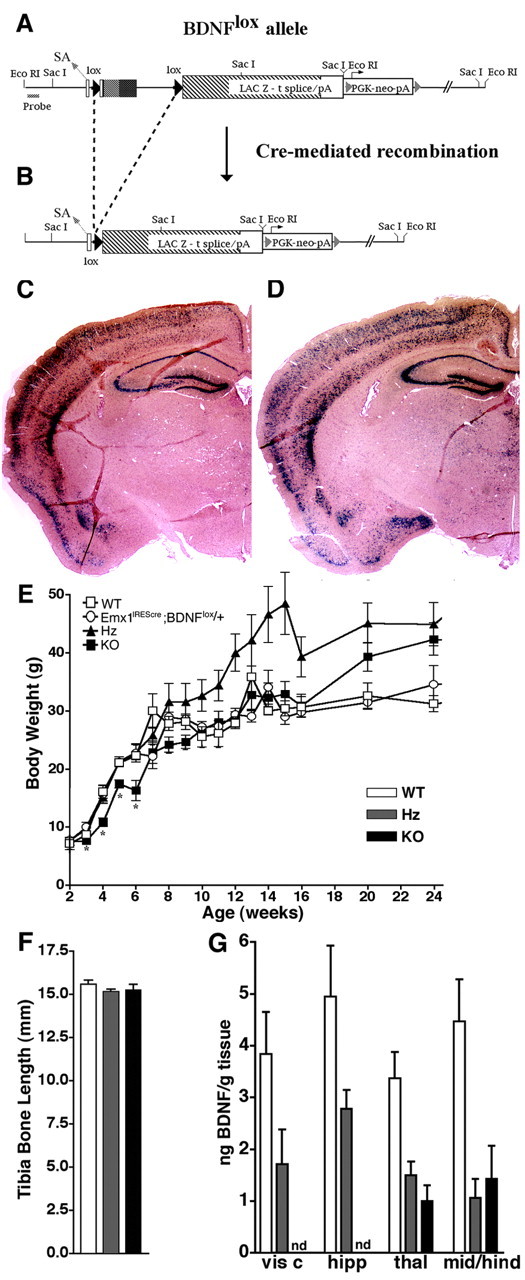
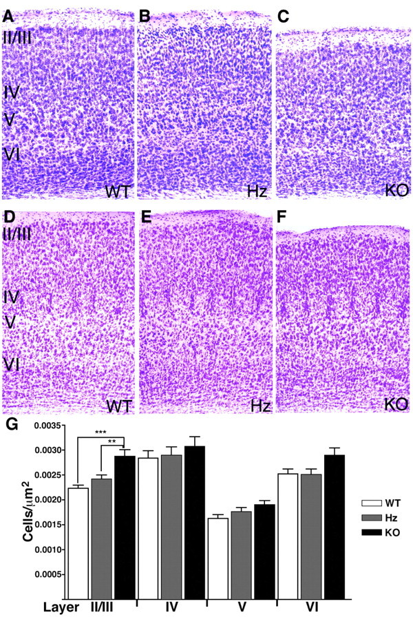
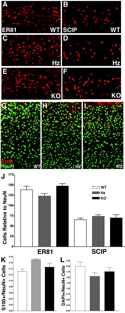
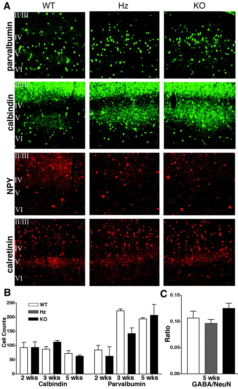
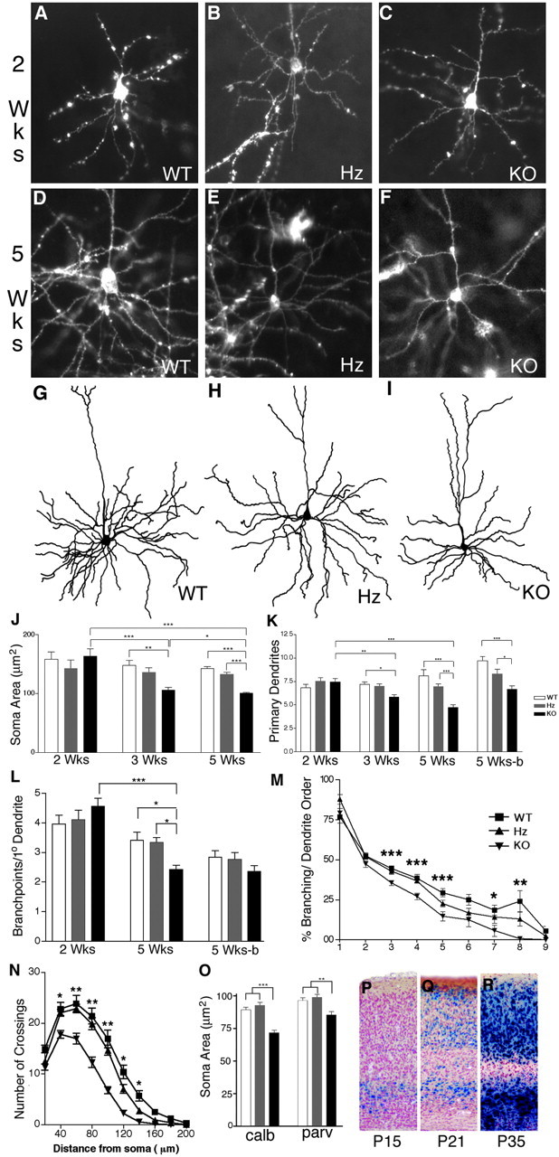
Brain-derived neurotrophic factor (BDNF) is thought to be involved in neuronal survival, migration, morphological and biochemical differentiation, and modulation of synaptic function in the CNS. In the rodent cortex, postnatal BDNF expression is initially low but subsequently increases to reach maximal levels around weaning. Thus, BDNF expression peaks at a time when both structural and functional maturation of cortical circuitry occurs. Although the function of BDNF has been probed using many approaches, its requirements during this phase of life have not previously been examined genetically. To test the in vivo requirements for BDNF during this important phase of development we generated early-onset forebrain-specific BDNF mutant mice. Although these mice undergo forebrain-restricted deletion of BDNF by Cre-mediated recombination during embryogenesis, they are healthy, and we did not detect the loss of specific cortical excitatory or inhibitory neurons. However, the neocortex of 5-week-old mice was thinner, attributable at least partly to neuronal shrinkage. Importantly, although visual cortical layer 2/3 neurons in the mutants initially developed normal dendrite structure, dendritic retraction became apparent by 3 weeks of age. Thus, our observations suggest that cortically expressed BDNF functions to support the maintenance of cortical neuron size and dendrite structure rather than the initial development of these features. This is consistent with a role for BDNF in stabilizing the "survival" of circuitry during the phase of activity-dependent reorganization of cortical connectivity.


Figures

Figure 1.
Figure 1.
Generation of forebrain-restricted BDNF mutant mice. A, Diagram of the BDNFlox allele. Exon V, the BDNF coding exon, is shown schematically at the top [open bar, 5′-UTR; shaded bar, pro region (a portion of the BDNF procursor protein removed by proteolytic processing); filled bar, mature hormone region], flanked by lox sites (filled triangles), and followed by lacZ (hatched bar), an SV40 intron-polyA (open bar), and an FRT-flanked PGK neomycin (open bar with triangles). Location of the splice acceptor (SA) is denoted by a narrow. B, Diagram of the BDNFlox allele after Cre-mediated recombination. BDNF-coding sequences in exon V are excised and lacZ is brought downstream of the splice acceptor site and under control of BDNF promoters. C, X-gal stained 10 μm coronal section through Emx1IREScre; BDNFlox/+ brain. D, X-gal stained 10 μm coronal section through BDNFlacZ/+ brain. E, Growth of wild-type (WT), Emx1IREScre; BDNFlox/+, BDNFneo/+ heterozygous (Hz), and Emx-BDNFKO mice (KO). Emx-BDNFKO mice weigh significantly less at 3, 4, 5, and 6 weeks of age. *p < 0.05. F, Tibia bone length measured for 5-week-old mice (n >10 mice per genotype). No significant differences were detected between genotypes. G, BDNF protein quantitated by ELISA, expressed as nanograms of BDNF protein per gram of wet tissue, in extracts of brain structures from 5-week-old mice (n = 3 mice per genotype). BDNF was not detected (nd) in the visual cortex and hippocampus of Emx-BDNFKO mice. visc, Visual cortex; hipp, hippocampus; thal, thalamus; mid/hind, midbrain/hindbrain.
Figure 2.
Figure 2.
Cortical thinning in Emx-BDNFKO mice. Cresyl violet-stained 50 μm coronal sections of visual (A-C) and somatosensory (D-F) cortices from 5-week-old WT, Hz, and KO mice. Cortical thickness is reduced in KO mice, but the cytoarchitecture appears generally normal and barrels are apparent in somatosensory cortex (D-F). G, Neuronal density in visual cortex of 5-week-old mice (n = 3 mice per genotype). Neurons are distributed more densely in layer II/III of KO mice (p < 0.05).
Figure 3.
Figure 3.
Absence of detectable neuronal or glial losses at 5 weeks of age. A-F, (20 μm) of layer V of primary visual cortex processed for ER81 (A, C, E) or SCIP (B, D, F) immunocytochemistry. G-I, Coronal sections primary visual cortex sections (10 μm) processed for S100 and NeuN immunoreactivity. Quantitation of ER81 and SCIP-positive layer V neurons relative to NeuN-positive cells (J), ratio of S-100-positive astrocytes to NeuN-positive neurons (K), and the ratio of total cells to NeuN-labeled neurons (L) revealed no differences between the genotypes (p > 0.05, n = 3 mice per genotype).
Figure 4.
Figure 4.
Apparently normal biochemical differentiation of cortical inhibitory neurons. A, Coronal sections (40 μm) of primary visual cortex from 3-week-old mice processed for parvalbumin, calbindin, NPY, and calretinin immunoreactivity revealed no gross differences in expression pattern. B, Quantification of calbindin- and parvalbumin-positive neurons from primary visual cortex of 2-, 3-, and 5-week-old WT and KO mice revealed no differences between the genotypes (p > 0.05, n = 3 mice per genotype). C, The ratio of GABA+/NeuN+ neurons is not significantly different at 5 weeks of age (p > 0.05, n = 3 mice per genotype).
Figure 5.
Figure 5.
BDNF is required for the maintenance of layer II/III pyramidal neuron morphology. Examples of DiI labeled visual cortical layer II/III pyramidal neurons from 2-week-old (A-C) and 5-week-old (D-F) mice and biocytin-filled and camera lucida-reconstructed layer II/III visual cortical pyramidal neurons from 5-week-old mice (G-I). J, Soma area of layer II/III neurons at 2, 3, and 5 weeks of age (n = 3 mice per genotype, 15 cells per mouse). K, Number of primary dendrites from 2-, 3-, and 5-week-old mice. *p < 0.05; **p < 0.01; n = 3 mice per genotype, 15 cells per mouse. L, Number of branch points per primary dendrites for 2- and 5-week-old mice (n = 3 mice per genotype, 15 cells per mouse). M, Number of branches per dendrite order for 2- and 5-week-old mice. *p < 0.05; **p < 0.01; n = 3 mice per genotype, 15 cells per mouse. H, Modified Sholl analysis of layer II/III pyramidal neurons from 5-week-old mice. *p < 0.05; **p < 0.01; n = 3 mice per genotype, 7 cells per mouse. Data in J-L were obtained from neurons visualized through diolistic labeling, with the exception that biocytin-filled and camera lucida-reconstructed neurons were used for some of the 5-week-old animals for comparison (-b). Data in M and N were obtained from biocytin-filled and camera lucida-reconstructed neurons. O, Soma areas measured from calbindin (calb) and parvalbumin (parv)-positive cortical neurons from 3-week-old mice revealed a significant difference between Emx-BDNFKO and control genotypes. **p < 0.01; ***p < 0.01; n = 3 mice per genotype, 55-75 cells per mouse. P-R, BDNF expression during postnatal cortical development as detected by X-gal staining of 40 μm sections from BDNFlacZ/+ mice. Postnatal ages are indicated at the bottom.

Similar articles

Cited by

References

    1. Akaneya Y, Tsumoto T, Hatanaka H ( 1996) Brain-derived neurotrophic factor blocks long-term depression in rat visual cortex. J Neurophysiol 76: 4198-4201. - PubMed
    1. Akaneya Y, Tsumoto T, Kinoshita S, Hatanaka H ( 1997) Brain-derived neurotrophic factor enhances long-term potentiation in rat visual cortex. J Neurosci 17: 6707-6716. - PMC - PubMed
    1. Alcantara S, Frisen J, del Rio JA, Soriano E, Barbacid M, Silos-Santiago I ( 1997) TrkB signaling is required for postnatal survival of CNS neurons and protects hippocampal and motor neurons from axotomy-induced cell death. J Neurosci 17: 3623-3633. - PMC - PubMed
    1. Antonini A, Fagiolini M, Stryker MP ( 1999) Anatomical correlates of functional plasticity in mouse visual cortex. J Neurosci 19: 4388-4406. - PMC - PubMed
    1. Araki T, Yamada M, Ohnishi H, Sano SI, Hatanaka H ( 2000) BIT/SHPS-1 enhances brain-derived neurotrophic factor-promoted neuronal survival in cultured cerebral cortical neurons. J Neurochem 75: 1502-1510. - PubMed

Publication types

MeSH terms