. 2003 Jul 30;23(17):6876-83.
doi: 10.1523/JNEUROSCI.23-17-06876.2003.

Development and subunit composition of synaptic NMDA receptors in the amygdala: NR2B synapses in the adult central amygdala

Affiliations

Development and subunit composition of synaptic NMDA receptors in the amygdala: NR2B synapses in the adult central amygdala

Mikel Lopez de Armentia et al. J Neurosci. .

Abstract

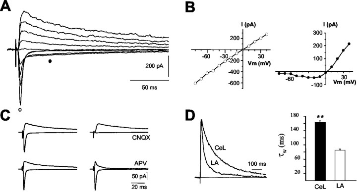
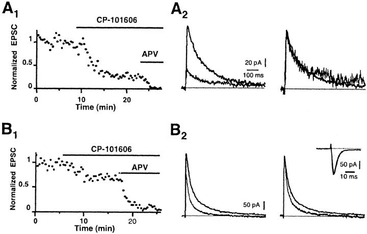
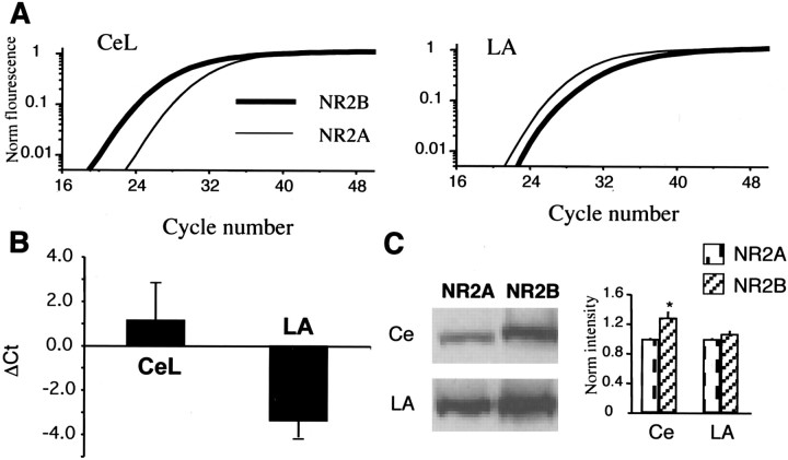
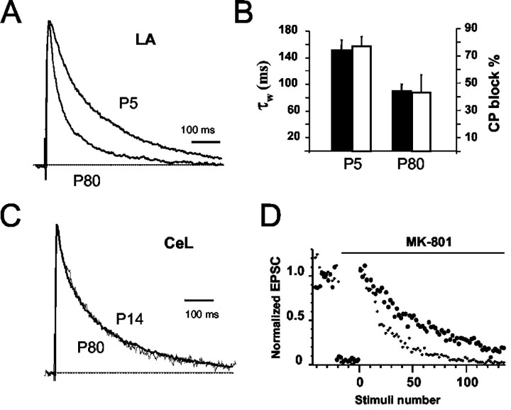
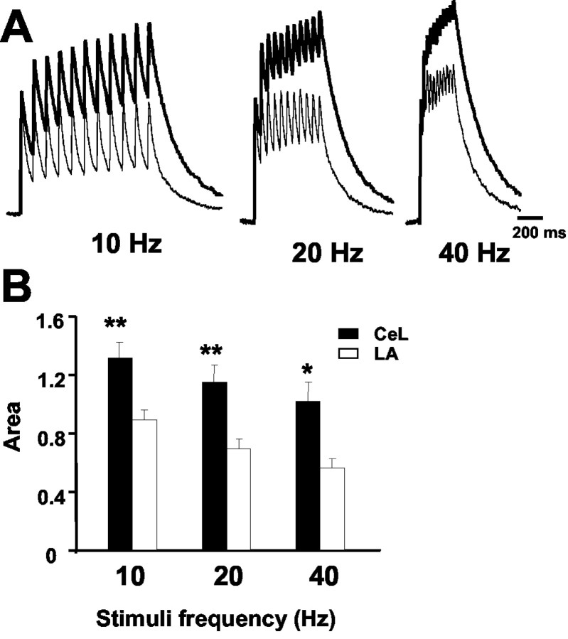
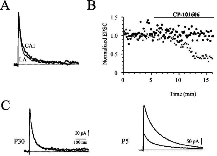
NMDA receptors are well known to play an important role in synaptic development and plasticity. Functional NMDA receptors are heteromultimers thought to contain two NR1 subunits and two or three NR2 subunits. In central neurons, NMDA receptors at immature glutamatergic synapses contain NR2B subunits and are largely replaced by NR2A subunits with development. At mature synapses, NMDA receptors are thought to be multimers that contain either NR1/NR2A or NR1/NR2A/NR2B subunits, whereas receptors that contain only NR1/NR2B subunits are extrasynaptic. Here, we have studied the properties of NMDA receptors at glutamatergic synapses in the lateral and central amygdala. We find that NMDA receptor-mediated synaptic currents in the central amygdala in both immature and mature synapses have slow kinetics and are substantially blocked by the NR2B-selective antagonists (1S, 2S)-1-(4-hydroxyphenyl)-2-(4-hydroxy-4-phenylpiperidino)-1-propano and ifenprodil, indicating that there is no developmental change in subunit composition. In contrast, at synapses on pyramidal neurons in the lateral amygdala, whereas NMDA EPSCs at immature synapses are slow and blocked by NR2B-selective antagonists, at mature synapses their kinetics are faster and markedly less sensitive to NR2B-selective antagonists, consistent with a change from NR2B to NR2A subunits. Using real-time PCR and Western blotting, we show that in adults the ratio of levels of NR2B to NR2A subunits is greater in the central amygdala than in the lateral amygdala. These results show that the subunit composition synaptic NMDA receptors in the lateral and central amygdala undergo distinct developmental changes.


Figures

Figure 1.
Figure 1.
NMDA receptor-mediated synaptic currents in the central nucleus have slow kinetics. A, Whole-cell recording from a neuron of the CeL. Synaptic currents were evoked at holding potentials of -80 to +40 mV in the presence of the GABA antagonist picrotoxin (100 μm). B, I-V of the fast component (open circles) and slow component (filled circles). The fast component has a linear I-V, whereas the slow component is blocked at negative holding potentials. Both components reversed near to 0 mV. C, The fast component was abolished by the AMPA/kainate antagonist CNQX (15 μm). The slow current present at deploarized potentials was blocked by the NMDA receptor antagonist d-APV (30 μm). D, Normalized NMDA receptor-mediated EPSCs recorded at +30 mV, in the presence of 10 μm CNQX, from a CeL neuron and LA pyramidal neuron, have been superimposed. Note that the current decay is much faster in LA neurons. The histogram shows the average weighted decay time constants (see “Materials and Methods”) of the NMDA receptor-mediated EPSC in CeL and LA neurons. All recordings were from animals P21-P30.
Figure 2.
Figure 2.
NMDA receptors at synapses in CeA neurons contain NR2B subunits. A1, Normalized amplitude of NMDA EPSCs recorded from a neuron in the CeA in the presence of CNQX and picrotoxin. Application of the NR2B-specific antagonist CP-101,606 (5 μm) largely blocked the EPSC. The remaining EPSC in CP-101,606 is blocked by d-APV (30 μm), showing that it is mediated by NMDA receptors. A2, NMDA EPSCs in control ringer and in the presence of CP-101,606 have been superimposed (left traces). When the EPSC in the presence of the NR2B antagonist is normalized to the control EPSC, there is no change in kinetics showing that it is also mediated by receptors containing NR2B subunits. B, Effect of CP-101,606 on the NMDA receptor-mediated EPSC in a pyramidal neuron in the LA. B1, Normalized amplitude of NMDA EPSCs recorded from a neuron in the LA in the presence of CNQX and picrotoxin. Application of 5 μm CP-101,606 partially blocks the EPSC, and the remaining EPSC is blocked by d-APV (30 μm), confirming that it is mediated by NMDA receptors. B2, EPSCs in control and CP-101,606 are shown superimposed. Normalizing the traces shows that the slow component of the EPSC is selectively blocked by CP-101,606 in LA neurons. The inset shows AMPA receptor-mediated EPSCs recorded at -70 mV from a neuron in the CeL before and after application of CP-101,606. CP-101,606 has no presynaptic effect on transmitter release at these synapses. All recordings were from animals P21-P30.
Figure 3.
Figure 3.
NR2B subunits are expressed at higher levels in the central nucleus as compared with the LA in animals P21-P30. A, Real-time PCR fluorescence is plotted against cycle number using primers against the NR2A subunit (thin line) and NR2B subunit (thick line). In the central amygdala (Ce), the fluorescence change for NR2B subunits occurs before that for NR2A subunits, whereas in the LA, fluorescence for 2A subunits rises before that for NR2B subunits. B, Ratio of threshold cycle (Ct) between NR2A and NR2B subunits for the Ce and LA. In each case, tissue from the Ce and LA were run in parallel. C, Western blot of protein levels for NR2A and NR2B subunits in the Ce and LA. The bar graph on the right plots the intensity levels normalized to that for NR2A in the Ce and LA. NR2B levels are significantly higher than NR2A levels in the central nucleus.
Figure 4.
Figure 4.
NMDA receptors undergo a developmental change in the LA but not in the CeA. A, Normalized NMDA receptor-mediated EPSCs recorded from pyramidal neurons in the LA from animals at P5 and P80. B, Average weighted time constants (filled bars) and percentage block by CP-101,606 (open bars) for the two age groups in LA neurons. The slow decay of the EPSC in LA neurons at P5 is because of the presence of NR2B-containing subunits, as shown by the much larger block of the EPSC by CP-101,606. C, Normalized NMDA receptor-mediated EPSCs recorded from neurons in the CeL at P14 and P80. D, Release probability is higher at NR2B-containing synapses. Normalized amplitude of the NMDA receptor EPSC is plotted before and after application of 5 μm MK-801. Progressive block of the NMDA EPSC by MK-801 is faster at synapses in the central nucleus (small circles) as compared with synapses in the LA (large circles).
Figure 5.
Figure 5.
Temporal summation of NMDA receptor-mediated EPSCs is larger in the CeL neurons as compared with LA neurons. A, NMDA receptor-mediated EPSCs recorded at +30 mV during repetitive stimulation at 10, 20, and 40 Hz. EPSCs recorded from CeL neurons (dark trace) and LA neurons (light traces) are superimposed. The first EPSC in the train has been normalized in each case. B, The area under the summed EPSCs, calculated after normalizing the first EPSC, has been plotted for inputs to neurons in CeL neurons (n = 6) and LA pyramidal neurons (n = 8). All recordings were from animals P21-P30.
Figure 6.
Figure 6.
NMDA receptors at Schaffer collateral to CA1 pyramidal neuron synapses are kinetically similar to those in LA neurons, but are pharmacologically different. A, Normalized NMDA EPSCs recorded in slices from P21-P30 rats from CA1 pyramidal neurons and an EPSC recorded in LA neurons have been superimposed (left). Average weighted time constants for NMDA receptor EPSCs from the LA and CA1 pyramidal neurons, showing that kinetically they are very similar. B, EPSCs in hippocampal neurons from P5 animals express higher levels of NR2B subunits. Block of the NMDA receptor-mediated EPSC by the NR2B-selective antagonist CP-101,606 (5 μm) at hippocampal CA1 synapses from animals at P5 (crosses) and at P30. NMDA EPSCs before and after application of CP-101,606 have been superimposed from P30 animals (top traces) and P5 animals (lower traces). CP-101,606 has no effect on EPSCs at mature synapses.

Similar articles

Cited by

References

    1. Behe P, Stern P, Wyllie DJ, Nassar M, Schoepfer R, Colquhoun D ( 1995) Determination of NMDA NR1 subunit copy number in recombinant NMDA receptors. Proc R Soc Lond B Biol Sci 262: 205-213. - PubMed
    1. Benveniste M, Mayer ML ( 1991) Kinetic analysis of antagonist action at N-methyl-d-aspartic acid receptors. Two binding sites each for glutamate and glycine. Biophys J 59: 560-573. - PMC - PubMed
    1. Blair HT, Schafe GE, Bauer EP, Rodrigues SM, LeDoux JE ( 2001) Synaptic plasticity in the lateral amygdala: a cellular hypothesis of fear conditioning. Learn Mem 8: 229-242. - PubMed
    1. Brimecombe JC, Boeckman FA, Aizenman E ( 1997) Functional consequences of NR2 subunit composition in single recombinant N-methyl-d-aspartate receptors. Proc Natl Acad Sci USA 94: 11019-11024. - PMC - PubMed
    1. Carmignoto G, Vicini S ( 1992) Activity-dependent decrease in NMDA receptor responses during development of the visual cortex. Science 258: 1007-1011. - PubMed

Publication types

MeSH terms

LinkOut - more resources